Key words
metabolic stress - time under tension - mitochondrial biogenesis
Introduction: Specific Adaptations to Divergent Stressors from Exercise
Introduction: Specific Adaptations to Divergent Stressors from Exercise
According to the principle of specificity, physiological adaptations reflect the
specific stress imposed on the body during various bouts of exercise [1]. Resistance training (RT) is associated with several
positive adaptations [2] such as increased muscular
endurance [3], muscular strength [4], power [5], sprint
speed [6], and agility [7]. These tangible measures of performance stem from adaptations that
occur at the nervous system and skeletal muscle. For example, common neurological
adaptations to RT include increased motor unit recruitment, faster transmission of
action potentials, increased rate coding, motor unit synchronization, and increased
surface area of the neural muscular junction [8]
[9]. At the skeletal muscle, RT increases fascicle
length, pennation angle, and hypertrophy (i.e., cross-sectional area of fibers
and/or muscle thickness) [10]
[11]
[12], which
potentially contribute to increased maximal force production [9]
[13]. These adaptations
are caused by the manipulation of several program variables including intensity,
volume, the order and exercises selected, rest intervals between sets, velocity of
contraction, and frequency [2]. Studies investigating
the role of the intensity and volume of RT have indicated that low-intensity,
high-volume RT and high-intensity, low-volume RT are effective to increase muscle
size and strength [14]
[15].
In contrast, aerobic training (AT) is associated with greater endurance capacity and
improvements in maximal oxygen uptake (VO2max), lactate threshold,
ventilatory threshold, and improved exercise economy [16]
[17]
[18].
Improved cardiovascular performance, stimulated by AT, stems from central and
peripheral adaptations. Central adaptations to AT generally include increased stroke
volume, cardiac output, and myocardial efficiency, meaning that the cardiovascular
system becomes more efficient at delivering oxygen to the exercising muscle [1]
[19]
[20]. Peripheral adaptations to AT include increased
capillary density, slow-twitch muscle fiber distribution, mitochondrial density, and
mitochondrial enzyme activity, meaning that the skeletal muscle becomes more
efficient at extracting oxygen from the blood stream and using it in the process to
synthesize adenosine triphosphate (ATP) via oxidative phosphorylation [1]
[19]
[20]. Research has demonstrated that high-volume,
low-intensity (i.e. long slow-distance) and low-volume, high-intensity AT (i.e.
high-intensity interval training or sprint interval training) are both capable of
stimulating central and peripheral aerobic adaptations [20]
[21]
[22].
As it pertains to skeletal muscle physiology at the molecular level, adaptations to
AT and RT are often viewed through a dichotomous lens in which they are not
compatible [23]
[24]
[25]. In particular, the mechanical tension imposed by
RT activates an unidentified protein kinase that upregulates the mammalian target
of
rapamycin (mTOR) by inhibiting the inhibitor of mTOR, tuberous sclerosis complex 2
(TSC-2) [25]
[26]. This
process (i.e., mechanotransduction) initiates acute protein translation which
eventually leads to long-term skeletal muscle hypertrophy [25]
[26]. In contrast, the metabolic stress
associated with AT upregulates various protein kinases that stimulate mitochondrial
biogenesis through activation of the peroxisome proliferator-activated receptor
gamma coactivator 1-alpha (PGC-1α) [27], that
is recognized as the master regulator of mitochondrial biogenesis [28]. Although chronic adaptations depend on training
status and the type of exercise performed, there is some evidence that AT can
stimulate RT adaptations and vice versa [29]
[30], which means there is cross-over between these
signaling pathways. Specifically, some research has demonstrated that AT elicits
significant skeletal muscle hypertrophy [31]
[32], and others have determined that RT can stimulate
increased skeletal muscle oxidative capacity [33] and
markers of mitochondrial biogenesis [34]. The latter
phenomenon is of particular interest in the current review because the potential RT
variables (i.e., volume, intensity, and tempo) that lead to aerobic adaptations are
not well understood.
Previously, Steele et al. [35] concluded that RT can
stimulate several AT adaptations such as increased cardiac output and
VO2max when sets are performed to momentary muscular failure. As it
pertains to skeletal muscle adaptations, the effects of RT on mitochondrial
biogenesis [36] and mitochondrial volume [37] have also recently been reviewed and a similar
conclusion was reached: Low-intensity, high-volume RT (e.g., high time under tension
– TUT) is likely a stronger stimulus for traditionally aerobic adaptations
compared to high-intensity, low-volume RT (e.g., low-TUT) [36]
[37]. However, these review papers did
not discuss the mechanisms by which high-TUT RT elicits such adaptations or provide
a general overview of acute and chronic adaptations to high-TUT training modalities.
Hence, the purpose of the current review is to detail the mechanisms of
exercise-induced mitochondrial biogenesis, provide a case for why high-TUT can
stimulate this pathway, and highlight what is known about three specific types of
high-TUT RT: Slow repetition tempo RT, traditional low-intensity RT, and drop-set
RT. Specifically, the acute physiological responses and long-term muscular
adaptations will be reviewed for each style of training.
Mitochondrial Biogenesis: The Central Role of PGC-1α Activation
Mitochondrial Biogenesis: The Central Role of PGC-1α Activation
Mitochondrial biogenesis is the synthesis of new reticular components that increase
mitochondrial volume (i.e., increased quantity) and the activity of enzymes within
the mitochondria (i.e., increased quality). Although other signaling cascades
contribute to mitochondrial biogenesis [38],
PGC-1α is considered to be the master regulator and key influencer for
aerobic adaptations and oxidative phenotypes [1]
[19]
[28]. In fact,
PGC-1α has been implicated in the regulation of skeletal muscle
mitochondrial biogenesis as muscle specific deletion of PGC-1α results in
attenuated mitochondrial biogenesis in response to physical training [39]. During exercise, repeated muscular contractions
lead to an increase in several contractile-induced stressors such as AMP/ATP
ratio, reactive oxygen species (ROS), intracellular Ca2+,
lactate, ischemia, and decreased energy availability [1]
[19]
[22]
[25]
[27].
These signals activate several protein kinases such as calcium/calmodulin
protein kinase II (CaMKII), p38 mitogen-activated protein kinase (p38MAPK), and
AMP-activated protein kinase (AMPK), which activate downstream transcription factors
and co-activators to increase the expression of mitochondrial proteins [1]
[19]
[22]
[25]
[27]. Specifically, CaMKII, p38MAPK, and AMPK directly
stimulate mitochondrial biogenesis by phosphorylating PGC-1α, causing it to
translocate to the nucleus [25]
[27]
[30]. Moreover, AMPK,
lactate, and NAD+activate NAD+-dependent
deacetylase family of sirtuins (SIRT), which controls metabolic flux through the
citric acid cycle and has been implicated in mitochondrial biogenesis by regulating
PGC1- α activity through its deacetylase activity [40]. Tumor suppressor protein 53 (p53) is also activated by AMPK and
p38MAPK, and it exerts regulatory effects on transcription factors and mitochondrial
content [1]
[19]
[27]. In short, exercise-induced metabolic stress and
increased skeletal muscle energy turnover activate upstream regulators of
PGC-1α, which converge on and phosphorylate PGC-1α, allowing for its
translocation.
When PGC-1α translocates to the nucleus, it activates several transcription
factors such as nuclear respiratory factors one and two (NRF-1 and -2), which
increase the transcription of PGC-1α, cytochrome c oxidase subunits, and
mitochondrial transcription factor A (TFAM) [1]
[19]
[24]
[27]. TFAM modulates mitochondrial biogenesis by
affecting mitochondrial DNA transcription and replication [41]. There is also evidence that PGC-1α influences angiogenesis
by upregulating the activity of vascular endothelial growth factor (VEGF) [18]
[42]
[43]. Thus, the repeated upregulation of PGC-1α
leads to post-exercise transcription of genes involved in mitochondrial biogenesis
and angiogenesis, which eventually leads to peripheral physiological adaptations.
For example, data from acute studies suggest that high-intensity interval training
(HIIT) elicits significant metabolic stress and the activation of PGC-1α,
which leads to increased levels of gene transcripts regulated by PGC-1α
[27]
[44]
[45]. When training bouts are repeated, HIIT leads to
increased oxygen uptake, mitochondrial volume, mitochondrial enzyme activity, and
capillary density [18]
[21]
[22].
Some have compared HIIT to RT because they are both characterized by brief periods
of
high-energy turnover interspersed by periods of rest [34]
[46]
[47].
Considering that energy depletion (e.g., reduced ATP and CrP) and metabolic stress
increase with the number of repetitions completed per set [48]
[49], we speculate that sets of RT with
high-TUT may stimulate the activation of upstream modulators of PGC-1α
similar to HIIT, leading to greater PGC-1α mRNA response, and ultimately to
enhanced mitochondrial biogenesis. For example, a set of RT performed for 5
repetitions with a 3-second tempo (e.g., 2:1 seconds) and high intensity
(e.g., 90% of 1-RM) would have a TUT of 15 seconds while a set of RT
performed for 20 repetitions with the same tempo but low intensity (e.g.,
50% of 1-RM) would have a TUT of 60 seconds. Gronneback and Vissing
[36] suggested in a recent review that the latter
style of RT (i.e., high-TUT) would have a positive effect on mitochondrial
biogenesis because it stimulates greater turnover rate of ATP, metabolic stress, and
tissue deoxygenation compared to low-TUT RT. More recently, Parry et al. [37] stated that future research should be done to
assess the effect of high-intensity (i.e., low-TUT) and low-intensity (i.e.,
high-TUT) RT on mitochondrial biogenesis, and hypothesized that the latter would
have a greater effect due to greater metabolic perturbations (i.e., higher blood
lactate). We submit that this hypothesis is interesting and the relationship between
blood lactate and mitochondrial biogenesis is worth further discussion.
Upregulating PGC-1α: The Potential Role of Lactate
Upregulating PGC-1α: The Potential Role of Lactate
Mechanistic studies in cell cultures and rodents have provided strong evidence that
lactate is involved in mitochondrial adaptations. Specifically, it has been
demonstrated that incubation of L6 cells with lactate increased mRNA expression of
PGC-1α [50], and similar results were achieved
in vivo by lactate intraperitoneal administration in mice [51]. Interestingly, attenuation of the increase in lactate during
exercise by administration of dichloroacetate, an activator of pyruvate
dehydrogenase, reduced HIIT-induced increases in mitochondrial enzyme content in
mouse skeletal muscles [52], implicating
exercise-induced lactate production in mitochondrial adaptations. Moreover,
Takahashi et al. [53] demonstrated that 3-week lactate
intraperitoneal administration increased mitochondrial enzyme activity (e.g.,
citrate synthase, 3-hydroxyacyl CoA dehydrogenase, and cytochrome c oxidase), and
showed that lactate administration prior to endurance exercise training enhanced
training-induced mitochondrial enzyme activity in the skeletal muscle [53]. Later, the same group showed that four weeks of
oral lactate administration+exercise increased cytochrome c oxidase activity
in skeletal muscle more than exercise alone in mice [54]. Finally, it was reported that chronic intramuscular lactate
treatment increased PGC-1α and citrate synthase protein content in the
gastrocnemius of rats [55]. Altogether, these findings
suggest that lactate increases mitochondrial enzymes through PGC-1α
activation, and that exercise-induced mitochondrial adaptations are related to
lactate production.
Although the precise pathways activated by lactate to induce PGC-1α-mediated
mitochondrial adaptations are still under investigation, it has been reported that
lactate injection can increase AMPK activity in the soleus muscle in mice [56]. Because AMPK is an upstream activator of
PGC-1α [27], it could be involved in
lactate-induced PGC-1α activation and mitochondrial adaptations. Conversely,
in vitro [57] and in vivo [58] evidence shows that when ROS production is blunted,
contraction-induced PGC-1α response is impaired, suggesting that ROS is an
important mediator of exercise-induced PGC-1α activation. Although it has
been suggested that the lactate upregulation of PGC-1α is mediated by ROS
[59], there is currently no direct evidence to
confirm this hypothesis. However, it was recently reported that lactate increased
ROS generation in a dose-dependent manner in skeletal muscle [60]. Therefore, we speculate that lactate accumulation
during exercise increases ROS production, which would lead to a ROS-mediated
PGC-1α activation culminating in mitochondrial biogenesis. Finally, lactate
might also affect mitochondrial biogenesis in an autocrine and/or paracrine
fashion. It has been reported that a selective receptor for lactate, called
G-protein-coupled receptor 81 (GPR81), exists in various tissues, including skeletal
muscle [61]
[62].
Interestingly, silencing of GRP81 in lactate-treated cancer cells did not increase
PGC-1α mRNA expression, in contrast to the increase observed in control
cells [63], suggesting that lactate induces
PGC-1α gene transcription by GRP81 activation. Whether the lactate-GRP81
pathway plays a role in exercise-induced mitochondrial biogenesis requires further
investigation.
These mechanistic studies suggest that lactate may be implicated as a signaling
molecule that mediates mitochondrial adaptations through PGC-1α activation,
but research in human subjects is equivocal. For instance, greater lactate
accumulation during cycle ergometry (i.e., repeated supramaximal sprints) has been
associated with higher exercise-induced phosphorylation of CaMKII and p38 MAPK,
along with higher PGC-1α mRNA response [64].
Although a direct cause-and-effect relationship between lactate accumulation and
PGC-1α activation cannot be established with their study design, the authors
speculated that greater metabolic stress (i.e., higher blood lactate) is related to
greater activation of the PGC-1α mRNA response [64]. In contrast, Moberg et al. [65]
recently reported that higher levels of muscle lactate did not facilitate increased
mRNA encoding of PGC-1α following RT with and without preceding lower-body
cycle ergometry. However, these results should be interpreted with caution because
both conditions elicited a robust muscle lactate response (10.8 vs.
13.5 mmol/L) which did not allow for a dose-response assessment
between lactate and PGC-1α mRNA. In other words, 10.8 and
13.5 mmol/L elicited similar responses, but it is unknown if values
of 4, 6, or 8 mmol/L would have led to an inferior mRNA response
(i.e., there may exist a saturation point above which lactate does not affect
PGC-1α). Ultimately, it is difficult to isolate the effect of lactate on
mitochondrial biogenesis in human skeletal muscle during exercise because several
stressors/signals (e.g., lactate, energy turnover, hypoxia) converge on
PGC-1α where they elicit their effects concurrently.
Regardless of the exact mechanisms ([Fig. 1]), there
is evidence that lactate accumulation is associated with the activation of
mitochondrial biogenesis [50–55; 61–63] and that blood lactate has a
positive, linear relationship with TUT during sets of RT [66]
[67]
[68]
[69]
[70]
[71]. Moreover, Burd et al. [72] reported that higher-TUT RT (i.e., 30%
1-RM) resulted in greater sarcoplasmic protein synthesis (e.g., which includes
mitochondrial proteins) compared to lower-TUT RT (i.e., 90% 1-RM). Later,
the same researchers measured greater mitochondrial protein synthesis following a
bout of RT with high-TUT [73]. Thus, it is clear that
high-TUT RT leads to greater metabolic stress (i.e., greater lactate accumulation)
than low-TUT RT, but whether these styles of RT lead to divergent peripheral aerobic
adaptations deserves further discussion.
 Fig. 1 An overview of the proposed mechanism for how resistance
training (RT) with high time under tension (TUT) stimulates peripheral
aerobic adaptations by upregulating the peroxisome proliferator-activated
receptor gamma coactivator 1-alpha (PGC-1α) signaling cascade.
Slow-tempo, traditional, and drop-set are three applications of RT with
high-TUT that lead to high muscle and blood lactate concentrations.
Mechanistic studies in cells and rodents have demonstrated that lactate
increases activation of adenosine monophosphate activated protein kinase
(AMPK) and concentration of reactive oxygen species (ROS) which directly
upregulate the PGC-1α signaling cascade. Additionally, lactate may
stimulate PGC-1α by directly binding to its G-protein-coupled
receptor 81 (GPR81). Because high-TUT leads to greater lactate concentration
than low-TUT, and lactate has been implicated as a potential signaling
molecule in the PGC-1α signaling cascade, it is logical to suggest
that RT with high-TUT may facilitate aerobic adaptations through
PGC-1α.
Fig. 1 An overview of the proposed mechanism for how resistance
training (RT) with high time under tension (TUT) stimulates peripheral
aerobic adaptations by upregulating the peroxisome proliferator-activated
receptor gamma coactivator 1-alpha (PGC-1α) signaling cascade.
Slow-tempo, traditional, and drop-set are three applications of RT with
high-TUT that lead to high muscle and blood lactate concentrations.
Mechanistic studies in cells and rodents have demonstrated that lactate
increases activation of adenosine monophosphate activated protein kinase
(AMPK) and concentration of reactive oxygen species (ROS) which directly
upregulate the PGC-1α signaling cascade. Additionally, lactate may
stimulate PGC-1α by directly binding to its G-protein-coupled
receptor 81 (GPR81). Because high-TUT leads to greater lactate concentration
than low-TUT, and lactate has been implicated as a potential signaling
molecule in the PGC-1α signaling cascade, it is logical to suggest
that RT with high-TUT may facilitate aerobic adaptations through
PGC-1α.
Low vs. High-intensity RT: Effect on Peripheral Aerobic Adaptations
Low vs. High-intensity RT: Effect on Peripheral Aerobic Adaptations
Many studies that have compared low vs. high-intensity RT are comprised of blood flow
restriction (BFR) interventions. The effect of such training on angiogenesis and
mitochondrial biogenesis has recently been reviewed [18]. Because BFR training evokes high levels of ischemia, metabolic
stress, and hypoxia, its effect on muscle capillary density and oxidative metabolism
are particularly interesting. In fact, research on acute bouts of RT have
demonstrated that the addition of BFR to low-intensity RT decreased muscle
oxygenation [42], increased gene expression for
proteins involved in angiogenesis [42], and increased
markers of mitochondrial biogenesis [43]. Thus, it is
logical that low-intensity BFR training has stimulated increased capillarization
[74] and muscular endurance [75] when repeated for 3–8 weeks. As it pertains
to mitochondrial adaptations, one study has compared the effects of high-intensity
RT vs. low-intensity RT with BFR [76]. After six weeks
of training, data revealed that both BFR (4 sets, 30% of 1-RM) and high-load
RT (4 sets, 70% of 1-RM) had positive effects on mitochondrial protein
fractional synthetic rate and mitochondrial respiration with no differences between
groups. Citrate synthase increased only in the BFR group, but the difference did not
achieve statistical significance [76]. As it pertains
to aerobic adaptations, more research is needed to determine if low-intensity RT
with BFR is superior to traditional forms of high-intensity RT.
Regarding traditional RT (i.e., no BFR), only two studies have assessed the effect
of
low vs. high-intensity RT (i.e., without BFR) on capillarization, cellular
respiration, and markers of mitochondrial biogenesis and mitophagy. Holloway et al.
[77] compared the effect of low-repetition
(8–12 reps; 75–90% of 1-RM) vs. high-repetition
(20–25 reps; 30–50% of 1-RM) RT in resistance-trained men.
Data revealed that both programs had a positive effect on capillarization and
protein markers of vasodilation, implying that positive adaptations to the
microvasculature occurred irrespective of intensity and TUT [77]. Other findings were reported by Lim et al. [34] who compared the effect of three RT programs in untrained males:
30% of 1-RM to failure, 80% of 1-RM to failure, or 30% of
1-RM with work matched to the 80% of 1-RM group. Results indicated that
protein markers for mitochondrial biogenesis, mitochondrial capacity, and mitophagy
increased only in the group that trained with 30% of 1-RM to failure [34]. In their discussion, the authors speculated that
when sets of RT are performed with high-volume (i.e., high-TUT), the metabolic
stress incurred during the session leads to aerobic/oxidative adaptations
[34]. In short, the hypothesis that low-intensity,
high-TUT training would have a positive effect on peripheral aerobic adaptations is
logical, but the limited research done in this area remains inconclusive. Future
research should be done to assess this hypothesis and determine if training status
influences the effect of different intensities on such adaptations.
As displayed in [Fig. 2] it is now known that
hypertrophy occurs along a wide spectrum of intensities (30–90% of
1-RM) and corresponding rep-ranges (3–35 reps per set) [3]
[14]
[15]
[78]
[79]. Assuming a traditional 2:1 second
repetition tempo, this effective repetition range corresponds with
9–105 seconds of TUT per set. Because the effective range of TUT for
hypertrophy is wide, it behooves us to explore unique adaptations that occur at
extreme ends of the spectrum. For example, it is generally accepted that RT with
low-TUT (i.e., 80–95% of 1-RM) is superior for increasing maximal
strength [80]
[81] while
RT with high-TUT (i.e., 30–50% of 1-RM) is superior for muscular
endurance [3]
[78]. The
latter is the focus of the current review, and the following section will summarize
research on acute and chronic RT for three specific techniques that use high-TUT:
Slow-tempo, high-volume low-intensity, and drop-set training.
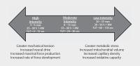 Fig. 2 A summary of the effective repetition range for hypertrophy
(3–35 reps) that emphasizes potential unique adaptations to
resistance training (RT) with low and high time under tension (TUT).
Assuming that a traditional repetition tempo is used (e.g.,
2:1 seconds), this repetition range also corresponds with a TUT of
9–105 seconds per set. Here, we submit that high-intensity
RT is associated with greater mechanical tension and increased strength
while low-intensity RT is associated with greater metabolic stress and
aerobic adaptations such as increased capillary density, mitochondrial
volume, and skeletal muscle oxidative capacity.
Fig. 2 A summary of the effective repetition range for hypertrophy
(3–35 reps) that emphasizes potential unique adaptations to
resistance training (RT) with low and high time under tension (TUT).
Assuming that a traditional repetition tempo is used (e.g.,
2:1 seconds), this repetition range also corresponds with a TUT of
9–105 seconds per set. Here, we submit that high-intensity
RT is associated with greater mechanical tension and increased strength
while low-intensity RT is associated with greater metabolic stress and
aerobic adaptations such as increased capillary density, mitochondrial
volume, and skeletal muscle oxidative capacity.
Applications of RT with High-TUT
Applications of RT with High-TUT
Slow Tempo Resistance Training
Repetition tempo, which is sometimes referred to as repetition duration, equals
the length of time that comprises the eccentric, isometric, and concentric
phases during one repetition of exercise [82]. For
example, a repetition with a three-second concentric phase, one-second isometric
pause, and three-second eccentric phase would be a seven-second tempo and would
be denoted as 3:1:3 sec [66]
[83]. As it pertains to muscular strength, in a
recent meta-analysis of 15 studies, Davies et al. [84] concluded that fast (e.g., eccentric
phase=1–3 seconds; concentric
phase=<1 second) and moderate-slow (e.g., eccentric
phase=1.7–3 seconds; concentric
phase=1.7–3 seconds) repetition tempos significantly
improve muscular strength. When considering skeletal muscle hypertrophy, in
another recent meta-analysis of 8 studies, Schoenfeld et al. [85] concluded that similar muscle growth occurred
along a wide repetition tempo spectrum (0.5–8 seconds) when sets
were performed to momentary muscle failure. Clearly, there is a wide range of
effective repetition tempos.
To the best of our knowledge, not much evidence is available regarding the effect
of repetition duration on muscular endurance and aerobic fitness. However, the
prospect of lengthening repetition duration to stimulate cardiovascular
adaptations is a noteworthy topic, because this will have a direct effect on the
TUT during sets of RT [86]. For example, a set of
12 repetitions with a 12-second duration (6:6 sec) would have a TUT of
144 seconds, while a set of 12 repetitions with a 2-second duration
(1:1 sec) would have a TUT of 24 seconds [73]. Although speculative, it is possible that sets
of RT with slower repetition tempos, and thereby longer TUT, have a positive
effect on peripheral aerobic adaptations because some research suggests that
metabolic stress (e.g., blood lactate) increases linearly with TUT [66]
[67]
[68]
[69]
[70]
[71]. Others have
shown that as TUT increases, muscle oxygenation decreases [66]
[83] while
mitochondrial protein synthesis increases [73].
Thus, the notion that slow-repetition, high-TUT RT can potentially stimulate
aerobic peripheral adaptations is a logical speculation.
Acute effect of repetition tempo on metabolic stress
There are several variations of repetition tempos that may influence
metabolic stress incurred during a bout of RT. Gentil et al. [89] compared the effect of four types of RT:
10-RM (2:2 second tempo), functional isometrics
(2:5:2 second tempo), vascular occlusion (20-second isometric
followed by repetitions with a 2:2 second tempo), and one super-slow
repetition (30:30 second tempo). The greatest blood lactate response
occurred in the functional isometric (4.5 mmol/L) and
vascular occlusion (4.2 mmol/L) conditions, and the authors
suggested that performing isometric pauses (5 or 20 seconds) had a
more profound effect on metabolic stress than overall TUT [89]. If their assertion is true, a
2:5:2 second tempo (i.e., 5 second isometric phase) would
increase blood lactate by more than a 6:3 second tempo (i.e., no
isometric phase) even though the repetition duration is the same (e.g.,
9 seconds). To date, this hypothesis has not been tested. In other
research, Mazzetti et al. [90] had ten
resistance-trained men perform lower-body RT under three conditions: Slow
(2:2 sec, 4×8 reps, 60% 1-RM), fast
(2:1 sec, 4×8 reps, 60% 1-RM), and heavy-fast
(2:1 sec, 6×4 reps, 80% 1-RM). Data indicated that
blood lactate increased linearly with TUT as slow
(TUT=32 sec) was greater than fast
(TUT=24 sec), which was greater than heavy-fast
(TUT=12 sec). However, it is difficult to provide definitive
conclusions from this study because subjective effort and proximity to
failure were not reported and the difference between tempos was narrow (3
vs. 4 sec).
With TUT matched at 36 seconds per set, Lacerda et al. [91] demonstrated that faster tempo repetitions
(3 seconds) increased blood lactate more than slower tempo
repetitions (6 seconds). Similar results were achieved by
Vargas-Molina et al. [92] when TUT was matched
at 60 seconds per set. This study improved upon the methods of
Lacerda et al. [91] because effort was matched
between conditions as every set was performed to momentary muscular failure.
Thus, there is agreement in the current literature that metabolic stress
increases with TUT [66]
[67]
[68]
[69]
[70]
[71]. Moreover, when TUT is matched, metabolic
stress is greater under conditions where more repetitions are performed per
set (e.g., 20 vs. 10 reps) and faster/traditional tempos (e.g., 3
vs. 6 sec) are used (91, 92). In the future, researchers should
emulate the design of Vargas-Molina et al. [92] by matching TUT and assessing the effect of several tempo
schemes on a variety of exercises (i.e., single vs. multiple joint, upper
vs. lower body). Furthermore, it would be beneficial to measure muscle
oxygenation during these exercises [66]
[83], and to include advanced biochemical
analysis (e.g., western blotting and immunohistochemistry) to measure
markers of mitochondrial biogenesis and angiogenesis.
Effect of tempo and TUT on long-term adaptations
Several recent systematic reviews and meta-analyses have conclusions positing
that significant hypertrophy and strength occur along a spectrum of fast,
traditional, slow, and super slow repetition tempos (e.g.,
0.5–10 seconds) [82]
[84]
[85].
Moreover, Tanimoto et al. [66] reported that
low-intensity RT with slow contractions (50% of 1-RM,
3:1:3 second tempo) and high-intensity RT with normal contractions
(80% of 1-RM, 1:1:1 second tempo) similarly increased
hypertrophy and muscular strength after training with the knee-extension
exercise. Years later, the same researchers reached similar conclusions when
applying these training styles to total body lifting with five exercises
[83]. Similar results were found when
these training styles were applied to elderly lifters [93], even when lower RT intensity was used
(30% of 1-RM) [94]. Together, these
studies demonstrate that the low-intensity, slow-tempo style of RT (i.e.,
7 seconds per repetition) can stimulate positive neuromuscular
adaptations when used in concert with low external loads corresponding to
30–60% of 1-RM.
Unfortunately, the researchers did not measure or report longitudinal
outcomes for muscular endurance or aerobic fitness in these studies [66]
[83]
[93]
[94]. However,
in their discussions, the authors made a case that the slow-tempo style of
lifting causes strong metabolic perturbation because the muscles
slowly/constantly occlude blood vessels, which causes deoxygenation
in a manner similar to BFR. This speculation warrants further investigation,
and the assessment of whether low-intensity slow-tempo training stimulates
increases in muscular endurance and aerobic fitness should be done.
Moreover, it will be important for future researchers to match the TUT
between conditions as the majority of the papers summarized in this section
compared very different TUT (i.e. 56 vs 24 seconds) conditions,
making it difficult to determine the effect of repetition tempos.
Traditional High-volume, Low-intensity RT
Resistance training adaptations (e.g., endurance, strength, and power) tend to be
specific to the combination of training variables used during a program. The
specificity of RT was best exemplified by Campos et al. [95] who reported that improvements in muscular endurance were
greatest in the high-repetition group (2 sets; 20–28 reps), increases in
muscular strength were greatest in the low-repetition group (4 sets; 3–5
reps), and hypertrophy only occurred in the low and intermediate-repetition
groups (3 sets; 9–11 reps) [95]. Although
their conclusions suggested that RT adaptations were largely specific to
intensity, more recent evidence suggests that improvements for hypertrophy,
strength, and power occur along a spectrum of 20–80% of 1-RM
[78]
[79].
Moreover, Schoenfeld (14), in a recent meta-analysis concluded that low
(<60% 1-RM) and high (>65% 1-RM) intensity RT
have similar and positive effects on muscular strength (9 studies,
n=251) and hypertrophy (8 studies, n=191). Thus, because
high-volume, low-intensity RT stimulates hypertrophy and strength, it is
intriguing to see if this style elicits unique benefits such as increased
muscular endurance and aerobic fitness.
Acute metabolic effects of high-volume, low-intensity RT
Lactate, an anaerobic by-product that is formed when pyruvate binds to two
hydrogen ions after glycolysis [92]
[96], is often used as a proxy measure of
metabolic stress during various styles of RT [89]
[90]. Rogatzki et al. [67] demonstrated that endurance-style RT (2
sets, 20 reps, 50% of 1-RM) elicited greater blood lactate response
than hypertrophy (3 sets, 10 reps, 70% of 1-RM) and strength (5
sets, 5 reps, 85% of 1-RM) RT during back squat exercise. Similarly,
da Silva et al. [68] showed a dose-response
relationship between TUT and blood lactate concentration during 8, 10, and
12 RM training on the bench press. In addition to lactate, transient
increases in the “anabolic hormones”, such as growth hormone
(GH), insulin growth factor 1 (IGF-1), and testosterone [97], have been indicated as proxy markers of
metabolic stress during RT [98]. Fink et al.
[87] demonstrated that training with
40% of 1-RM significantly increased IGF-1 and GH after training with
bench press and back squat. Compared to training with 8 RM, the same
researchers reported that GH concentration was only elevated after training
with 20 RM [88].
The preponderance of research summarized above suggests that metabolic stress
increases as TUT and repetition number increase, especially when it is
measured via blood lactate. However, there is a paucity of research that has
compared the acute effect of different repetition ranges (e.g., 10-RM vs.
20-RM) and TUT (e.g., 30 vs. 60 seconds) on markers of metabolic
stress, muscle oxygenation, and mitochondrial biogenesis during RT. Future
researchers could design studies to match proximity to failure and
repetition tempo (e.g., 2:1 sec), and have participants perform a
lower-body exercise (e.g., belt squat) with external loads of 10-RM, 20-RM,
and 30-RM with corresponding TUT of 30, 60, and 90 seconds. As
suggested before, the researchers could measure muscle oxygenation, blood
lactate, and markers of mitochondrial biogenesis for all conditions.
Chronic effects of high-volume, low-intensity RT
Several studies have compared the effect of low vs. high intensity RT to
delineate if adaptations to RT are determined by the external load used. For
instance, Leger et al. [99] recruited 25
healthy, untrained males and randomly assigned them to low (4 sets,
3–5 repetitions) or high (2 sets, 20–28 repetitions per set)
volume RT. Their results showed that both training programs stimulated
increased muscular hypertrophy, endurance, and strength with no differences
between groups [85]. In a unilateral,
within-subject research design, Mitchell et al. [78] recruited 18 healthy, untrained males, and randomly assigned
their legs to one of three RT conditions: 3 sets with 30% 1-RM, 1
set with 80% 1-RM, and 3 sets with 80% 1-RM. Data indicated
that all groups significantly increased hypertrophy and strength.
Interestingly, for muscular endurance tasks, the 30% 1-RM condition,
participants increased the number of repetitions that they could perform
with 30% and 80% of their 1-RM. By contrast, neither
80% 1-RM condition increased participants’ repetition
performance with 30% of 1-RM [78].
Extending these research designs to trained subjects, Schoenfeld et al. [3] reported that low-load (25–35 reps,
30–50% of 1-RM) and high-load (8–12 reps,
70–80% of 1-RM) significantly increased hypertrophy and
strength. Of note, muscular endurance (i.e., repetitions to failure with
50% of 1-RM on bench press) only increased in the low-load group
[3]. Moreover, when compared to a group of
lifters who performed the same intensity for every training session
(8–10 RM), those who performed a daily undulating periodization plan
(2–4 RM, 8–10 RM, 25–35 RM) significantly increased
repetition performance with 50% of 1-RM on bench press [100]. This means that one weekly session of
low-intensity RT was enough to improve muscular endurance. Collectively, the
literature demonstrates that low-intensity, high-volume RT delivers several
adaptations to RT (e.g., endurance, hypertrophy, and strength), and future
research should be done to determine if such RT leads to increased oxidative
capacity (i.e., at the skeletal muscle) and improved aerobic performance. In
particular, it would be interesting to determine if there are sex
differences for such adaptations, as some research has demonstrated that
females tolerate metabolic stress better [101]
and can perform more repetitions at relative intensities compared to males
[102].
Drop-set Resistance Training
A brief research review by Schoenfeld and Grgic [103] identified drop-set RT as an effective way to accrue high levels
of training volume and to stimulate significant muscular adaptations in a short
amount of time. To perform a drop-set, the initial set of RT with a fixed
external load (e.g., 80% 1-RM) is performed to muscular failure. From
there, the load is immediately reduced by 20–25% (i.e., no rest)
and the lifter performs a subsequent set to muscular failure [103]. Although it is not strictly defined, the
authors suggest that two to three drops are performed during one drop-set, and
that the rest interval between drops should be kept to a minimum (i.e., just
long enough to adjust the load and ensure that the lifter is in a proper
starting position) [103]. When following these
guidelines, it is likely that a lifter will perform 20–30 consecutive
repetitions at intensities that correspond to 40–80% 1-RM in
just one set of exercise. Assuming a traditional 2:1 second eccentric to
concentric repetition tempo (i.e., three second contractions), this translates
to an approximate TUT of 60–90 seconds, which leads to
significant metabolic stress, ischemia, and hypoxia [103]. Although the authors presented drop-set training as a means to
evoke skeletal muscle hypertrophy [103], we submit
that this style of RT could be used to stimulate peripheral adaptations that are
typically associated with AT.
Acute metabolic effects of drop-set RT
Few studies have quantified the metabolic stress incurred during sessions of
drop-set RT. For example, Goto et al. [104]
demonstrated that the addition of one drop with 20, 30, or 50% of
1-RM after finishing a standard session of RT (5 sets, 90% of 1-RM)
significantly increased GH and blood lactate. Years later, the same research
team concluded that drop-set training stimulated significant decreases in
muscle oxygenation, especially in trained lifters who have greater muscle
thickness than their untrained counterparts [105]. Compared to straight-set training (i.e., no drop sets),
Fink et al. [106] reported that drop-set RT
elicited greater muscular swelling while increases in blood lactate were
similar. Considering that volume (reps x% of 1-RM) was similar
between groups (38.3 vs. 38.9 arbitrary units) the results from this study
suggest that both training styles elicited significant metabolic stress but
drop-set training did so in a more time-efficient manner (145 vs.
315 sec).
By examining the acute RT data summarized above, it is clear that drop-set
training delivers a strong metabolic load to the skeletal muscle as
indicated by increased blood lactate and decreased oxygenation during
exercise. As previously theorized, metabolic stress and ischemia may be key
factors that lead to peripheral adaptations that are intrinsic in AT such as
increased vascularization, blood flow, and mitochondrial biogenesis. Future
research should be done to evaluate the effect of drop-set RT on protein
markers of these adaptations while measuring lactate and muscle oxygenation
to help determine a cause-effect relationship between such training and
peripheral aerobic adaptations.
Chronic effects of drop-set RT
In a longitudinal design, Goto et al. [107]
concluded that strength training (5 sets, 90% of 1-RM) and strength
training with the addition of one drop set (25–35 repetitions with
40–50% of 1-RM) both led to significant increases in
endurance, strength, and rate of force development. However, the drop-set
group had significantly greater increases in 1-RM for leg press, maximal
isokinetic strength at a fast velocity (e.g., 300 degrees/second),
and muscular endurance, which was quantified as total work performed (load x
repetitions) during one set of knee-extension to failure with 30% of
maximal voluntary contraction [107]. Because
total training volume was not matched, it is difficult to conclude if the
differences between groups occurred strictly because of the metabolic stress
imposed by the drop-set condition. Others reported that drop-set and
traditional RT had similar effects on neuromuscular performance, especially
muscular endurance [108]. Ozaki et al. [109] revealed that high-intensity RT
(80% of 1-RM) and drop-set RT (1 set with 80% of 1-RM, 4
drop sets at 65, 50, 40, and 30% of 1-RM) elicited similar increases
in hypertrophy and strength while the drop-set condition led to better
endurance. It is important to note that the drop-set training delivered
significant adaptations despite the performance of ~1/3 of
the training volume (5,308 vs. 15,365 kg) with sessions that
required ~1/5 of the training time (2.1 vs.
11.6 minutes) compared to the low-load group [109].
Taken together, these studies support that drop-set RT is a time-efficient
strategy to promote meaningful neuromuscular adaptations, especially
muscular endurance. Indeed, when training volume is similar, it seems that
drop-sets do not confer additional adaptations when compared to traditional
forms of RT, but the concept of delivering such adaptations with shorter gym
sessions is important considering that time is reported to be a barrier to
exercise [103]
[110]. Future research should be done to determine if drop-set RT
leads to AT-like peripheral adaptations and if these adaptations lead to
improved aerobic exercise performance.
Conclusions and Directions for Future Research
Conclusions and Directions for Future Research
Traditionally, the physiological adaptations to AT and RT have been viewed through
a
dichotomous lens where AT stimulates the synthesis of mitochondrial proteins and RT
stimulates the synthesis of myofibrillar proteins. Recent research suggests
cross-over between these seemingly divergent training modalities as AT can cause RT
adaptations and vice versa. As it pertains to RT, we submit that low-intensity,
high-volume RT with high-TUT is an effective stimulus for peripheral aerobic
adaptations such as increased capillary density, mitochondrial volume, and oxidative
metabolism. This logical conjecture stems from the fact that RT with high-TUT leads
to significant metabolic perturbation, ischemia, and skeletal muscle hypoxia, which
upregulate signaling cascades for angiogenesis and mitochondrial biogenesis. More
research is needed to identify the exact mechanism, but the results from several
cell and rodent studies suggest that lactate may facilitate mitochondrial
adaptations through the PGC-1α signaling cascade. In other words, the stress
imposed by high-TUT RT reflects traditional forms of AT (i.e., HIIT), and the
specific adaptations to this stress may be similar between modalities. Research
shows that slow-tempo, traditional, and drop-set training are all effective
variations of high-TUT RT that increase skeletal muscle endurance, hypertrophy, and
strength. Based on acute data, these training modalities also evoke significant
metabolic stress and skeletal muscle hypoxia during exercise, and future research
can determine if this stress leads to aerobic adaptations.
Thus, there are several opportunities for future studies. Specifically, researchers
should better quantify the acute metabolic stress of high-TUT RT by measuring muscle
oxygenation, blood lactate, and upregulation of protein markers involved in
angiogenesis and mitochondrial biogenesis. Moreover, it would be interesting to
measure the chronic effect of high-TUT RT on aerobic capacity (e.g.,
VO2max) and aerobic performance (e.g., 5-km time trial). The influence of
training status is another possible area for research [30]. For example, it is likely that compared to trained lifters,
untrained counterparts would incur more metabolic stress during high-TUT RT, which
could potentially lead to superior long-term aerobic adaptations. It would be
interesting to apply this logic to resistance trained participants who typically
perform high-intensity, low-TUT RT. In other words, researchers can determine if
performing sets of RT with 60–90 seconds of TUT provides a novel,
aerobic stimulus for well-trained lifters who typically perform their RT sets with
10–30 seconds of TUT, and are, therefore, relatively untrained in
high-TUT RT [111]. Finally, in a recent review by
Schoenfeld et al. [112] it was concluded that the
repetition range for hypertrophy and strength is very wide and that unique
adaptations occur at either end of this spectrum ([Fig.
2]). At the low intensity end of the spectrum, it would be interesting to
follow the design of Lacerda et al. [91] and
Vargas-Molina et al. [92] by matching TUT (e.g.,
60 seconds) and proximity to failure (e.g., RPE of 8–9 out of 10)
while varying repetition tempo within the matched TUT (e.g., 20 reps at 2:1 vs. 10
reps at 4:2 vs. 6 reps at 6:4) to evaluate the true effect of repetition tempo on
aerobic (e.g., mitochondrial biogenesis) and resistance (e.g., strength)
adaptations. Similar study designs can be applied to higher-intensity RT with
shorter TUT (e.g., 30 seconds).
Ethical Standards
The authors confirm that the current review meets the ethical standards of the
International Journal of Sports Medicine as outlined by Harriss et al. [113].


