

Subscribe to RSS
DOI: 10.1055/a-1949-7641
Imaging Osteomyelitis: An Update
Bildgebung der Osteomyelitis: ein UpdateAuthors

Abstract
Background Hematogenous osteomyelitis has increased over the past quarter century in frequency, virulence, and degree of soft-tissue involvement, bringing about changes in clinical manifestations and management of the disease especially in children that should be reflected in the current imaging approach. Likewise, the global disease burden of diabetes has increased greatly in the same period, compounding the problem of ascertaining osteomyelitis in diabetic foot.
Method This article provides an updated overview of imaging findings in hematogenous and contiguous osteomyelitis based on the literature and our institutional experience, along with salient features of recent recommendations from expert groups on the diagnostic algorithms and reporting terminology.
Results and Conclusion Findings on radiography and especially magnetic resonance imaging (MRI) closely reflect pathophysiology in osteomyelitis, whereby the characteristic involvement of the metaphysis or metaphyseal-equivalents, the formation and subperiosteal extension of intramedullary pus collection, and the development of cloaca, sequestrum, and involucrum are all diagnostic clues. Non-enhancing foci within the medullary bone, the penumbra sign, intra- or extramedullary fat globules, and surrounding soft tissue inflammation or abscesses are among key MRI findings. Diabetic foot is a special condition with characteristic pathophysiologic and imaging features that suggest the likelihood of osteomyelitis and the main differential diagnostic consideration of acute on chronic neuropathic osteoarthropathy with or without osteomyelitis.
Key Points
-
Imaging closely reflects pathophysiology in hematogenous osteomyelitis.
-
Acute hematogenous osteomyelitis predominantly involves metaphyses and metaphyseal equivalent sites.
-
MRI clues for hematogenous osteomyelitis include central marrow non-enhancement, intra- or extramedullary fat globules, and the “penumbra” sign.
-
An increased fluid-sensitive MRI bone signal abutting a soft tissue ulcer, abscess, or sinus tract suggests a high probability of contact osteomyelitis.
Citation Format
-
Aydingoz U, Imaging Osteomyelitis: An Update. Fortschr Röntgenstr 2023; 195: 297 – 308
Zusammenfassung
Hintergrund Die hämatogene Osteomyelitis hat im letzten Vierteljahrhundert in Bezug auf Häufigkeit, Virulenz und Grad der Weichteilbeteiligung zugenommen, was insbesondere bei Kindern zu Veränderungen bei den klinischen Manifestationen und der Behandlung der Krankheit geführt hat. Dies sollte sich im aktuellen bildgebenden Ansatz widerspiegeln. Ebenso hat die globale Krankheitslast von Diabetes im gleichen Zeitraum stark zugenommen, was das Problem der Diagnostik der Osteomyelitis beim diabetischen Fuß noch verschärft.
Methode Dieser Artikel gibt, basierend auf der Literatur und unserer institutionellen Erfahrung, einen aktualisierten Überblick über die bildgebenden Befunde bei hämatogener und konsekutiver Osteomyelitis, sowie über die auffälligsten Merkmale nach den aktuellsten Empfehlungen von Expertengruppen zu diagnostischen Algorithmen und Befundterminologie.
Ergebnisse und Schlussfolgerungen Das Röntgenbild und insbesondere die Befunde der Magnetresonanztomografie (MRT) spiegeln die Pathophysiologie der Osteomyelitis sehr gut wider. Diagnostische Hinweise sind in diesem Fall die charakteristische Beteiligung der Metaphyse oder metaphysärer Äquivalente, die Bildung und subperiostale Ausdehnung intramedullärer Eiteransammlungen und die Entwicklung von Kloake, Sequester und Involucrum. Zu den wichtigsten MRT-Befunden gehören Herde mit Nicht-Enhancement innerhalb des Markraums, das Penumbra-Zeichen, intra- oder extramedulläre Fettkügelchen und Entzündung der umgebenden Weichteile oder Abszesse. Der diabetische Fuß ist ein spezielles Krankheitsbild mit charakteristischen pathophysiologischen und bildgebenden Merkmalen, die auf eine Osteomyelitis hindeuten und differentialdiagnostisch in erster Linie an eine akute oder chronische neuropathische Osteoarthropathie mit oder ohne Osteomyelitis denken lassen.
Kernaussagen
-
Die Bildgebung spiegelt die Pathophysiologie der hämatogenen Osteomyelitis genau wider.
-
Die akute hämatogene Osteomyelitis betrifft überwiegend Metaphysen und metaphysäre Äquivalente.
-
MRT-Anhaltspunkte für eine hämatogene Osteomyelitis sind Nicht-Enhancement des zentralen Knochenmarks, intra- oder extramedulläre Fettkügelchen und das Penumbra-Zeichen.
-
Ein erhöhtes flüssigkeitssensitives MRT-Knochensignal, das an ein Weichteilgeschwür, einen Abszess oder einen Sinustrakt angrenzt, spricht mit hoher Wahrscheinlichkeit für eine Kontakt-Osteomyelitis.
Introduction
Radiology plays several roles in the management of osteomyelitis, including initial diagnosis and follow-up [1] [2], image-guided sampling [3] [4] [5], and subperiosteal abscess drainage [6]. The role of imaging is well-established and indispensable in diagnosing osteomyelitis and tracking its progression or treatment response [1] [2], but is somewhat ambiguous in interventional radiologic procedures that are sometimes employed in its management [3] [4] [5] [6] [7].
Imaging closely reflects pathophysiology in osteomyelitis and has some unique, if not pathognomonic, findings that are helpful in suggesting the diagnosis. The purpose of this article is to provide an overview of the current landscape of the imaging diagnosis of osteomyelitis. An update on this topic is warranted based on several developments: First, hematogenous osteomyelitis has increased over the past quarter century in frequency, virulence, and degree of soft-tissue involvement, effecting changes in clinical manifestations and management of the disease, especially in children, that should be reflected in the current imaging approach [1]. Likewise, the global disease burden of diabetes has persistently and greatly increased in the same period, compounding the problem of ascertaining osteomyelitis in diabetic foot [8]. Second, there have been some changes in our understanding of the imaging appearances of osteomyelitis such as the observation that transphyseal spread of infection from the metaphysis to the epiphysis is more common than was classically suggested [9]. Third, new consensus or guideline documents prepared by experts came out in recent years, featuring updated recommendations on the appropriate use of imaging in diagnosing osteomyelitis and related terminology in radiology reports [10] [11] [12] [13]. In this article, first, I will review the pathophysiology of hematogenous osteomyelitis and how bone anatomy and imaging relate to it. Then, I will discuss several key imaging findings of osteomyelitis and its differential diagnosis along with a subsection focusing on diabetic foot. Throughout the narrative, I will point out the most salient parts of the recent recommendations on imaging diagnosis of osteomyelitis (which sometimes feature discrepancies between different expert groups) and updates on terminology to be used in radiology reports. Osteomyelitis in the spinal column and periprosthetic infections are beyond the scope of this review.
How imaging relates to bone anatomy and pathophysiology in osteomyelitis
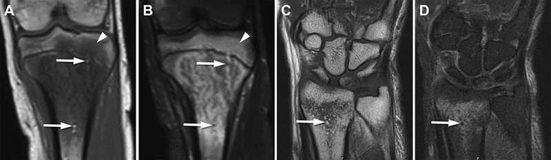
The hematogenous route is by far the most common means of spread in osteomyelitis ([Fig. 1A]). The other two modes involve direct contact as with a penetrating injury ([Fig. 1B]) (which is more common in hands and feet or following open fractures and surgery) or spread from an adjacent soft tissue infection ([Fig. 1C–E]), which is especially common in diabetic or otherwise neurologically impaired or bedridden patients.

The initial site of involvement is characteristically the metaphysis in children for hematogenous osteomyelitis owing to the rich vascularity at this location with vessels of slow flow and discontinuous endothelium, both of which facilitate a foothold for bacteria that settle and thrive there ([Fig. 2A], [Fig. 3A, B, E–H]). This is especially true during periods of rapid growth in childhood and, not surprisingly, acute hematogenous osteomyelitis is particularly common in children < 5 years of age (In contradistinction to infants and children, in whom the femur or tibia is involved in approximately half of all cases, adults are more likely to develop osteomyelitis in the spine, pelvis, or small bones of the hands and feet [13] [14]). Since the pelvis and lower extremities account for a vast majority of cases of osteomyelitis in children, it can be beneficial during MRI to first acquire coronal STIR images from the pelvis to the feet to locate the disease, especially in small children who usually have problems describing and pinpointing their symptoms [1] [14].


In addition to the metaphyses in long tubular bones, the so-called “metaphyseal equivalents”, which are located at the junction of bone and cartilage in skeletally immature flat or round bones, and the periphery of the epiphyseal (secondary) centers of ossification, have similar vascularity to the metaphysis of a long bone and are also particularly susceptible to osteomyelitis [1] [15]. These metaphyseal equivalents surround the triradiate cartilage, the ischiopubic synchondrosis, the sacroiliac joints, and the vertebral body endplates and are also located at the periphery of round bones such as the talus and calcaneus near its posterior apophysis ([Fig. 3C, D]). In these bones, hematogenous osteomyelitis tends to begin in the metaphyseal equivalent locations [1].
After the infection settles at the metaphysis (or its equivalents) in hematogenous osteomyelitis ([Fig. 2A, B]), the blood supply to the bone is blocked ([Fig. 2C]) as the infection progresses and pus accumulates, usually finding its way to under the periosteum all around the bone, thereby further blocking the vascular nourishment of the bone ([Fig. 2D]), which eventually results in areas of necrosis ([Fig. 2E]). An intraosseous abscess can form. According to the recent recommendations on MRI terminology for osteomyelitis from the Society of Skeletal Radiology (SSR), which is the premier musculoskeletal imaging association of North America-based radiologists, “intraosseous abscess” is an appropriate term for intraosseous fluid-signal cavities with a rim of peripheral enhancement, or in the presence of restricted diffusion or the penumbra sign (explained later in this article) if intravenous (i. v.) contrast is not administered [13]. “Brodie abscess” should be used for intraosseous abscesses in subacute (symptom duration 2–4 weeks) or chronic (symptom duration > 4 weeks) osteomyelitis ([Table 1]) [13].
|
Current term |
Recommended term |
Rationale |
|
Subperiosteal abscess |
Subperiosteal spread of infection |
It may be difficult to differentiate subperiosteal abscess from phlegmon |
|
Intraosseous abscess |
Intraosseous abscess or Brodie abscess |
“Brodie abscess” should be used for intraosseous abscesses in subacute or chronic osteomyelitis having a predilection for the ends of tubular bones |
|
Osteitis (in diabetic foot) |
Low likelihood of osteomyelitis |
“Osteitis” should be avoided in the case of concern regarding infection but still applies in non-infection cases like those due to inflammatory arthritis Vascular insufficiency may fail to produce marrow replacement or enhancement on T1-weighted images |
Besides subperiosteal extension, the infection may spread from its metaphyseal origin to the epiphysis, the adjacent joint space and soft tissues, and the diaphysis ([Fig. 2D]). The involvement of the neighboring joint space in the bone infection is via either epiphyseal or subperiosteal spread. The latter is possible when a metaphysis is intracapsular (i. e., the joint capsule inserts to a bone crossing over the growth plate such as in the femoral side of the hip joint or the radial side of the elbow joint). Over time, pus might escape from the bone by way of a “cloaca” ([Fig. 1A], [Fig. 2E], [Fig. 3C–G]), which in Latin means “sewer”. The dead bone tissue might be trapped within the infected bone (therefore called the “sequestrum”, which means “separated” or “isolated” in Latin) and enveloped by a new bone formation, which is called the “involucrum” ([Fig. 2E]). By virtue of its capability to show soft tissues and bone marrow in exquisite detail, MRI is the only imaging modality that shows this cascade of events ([Fig. 2], [3]) in its entirety – although some stages can also be vaguely identified on radiography. The novel zero echo-time (ZTE) sequence, which generates computed tomography- [CT] or radiography-like bone images on high-field (1.5 T or 3 T) MRI by picking up the scant signal from mineralized structures, can show sequestra more conspicuously than was ever possible on MRI before ([Fig. 4]) [16]. It is possible to make multiplanar reformations with the ZTE sequence, which is a virtually silent isotropic 3D technique that runs about 3–5 minutes [16]. We just published a comprehensive overview of the technique, optimization, applications, and pitfalls of this new MRI sequence [16].

The presence of abscesses in the subperiosteal space (and sometimes the adjoining soft tissues) can be so remarkable that in regions with complex anatomy, such as the pelvis, it may be challenging to ascertain the bony origin of infection [1]. In fact, pelvic osteomyelitis is associated with extensive soft tissue inflammation and abscesses (mostly involving the soft tissues overlying the infected bone – but not subperiosteal) in 55 % of cases, which may overshadow the original bone infection [17]. In such instances, it is helpful to remember the concept of metaphyseal equivalents and look for areas of bone infection at these sites [1]. The SSR recommends the term “subperiosteal spread of infection” instead of “subperiosteal abscess”, since it may be difficult to differentiate subperiosteal abscess from phlegmon [13] ([Table 1]).
The periosteum is loosely attached to the bone shaft in children. Pus from the metaphyseal infection can easily collect under the superficial, fibrous layer of the periosteum, which meets the bone cortex in a tight junction only near the perichondrium at the periphery of the physis ([Fig. 2C–E]), forming a “V” at the vertex ([Fig. 5]) [1]. When an abscess is encountered adjacent to a bone, it is important to bear in mind anatomic features like this, in order to distinguish whether the abscess is subperiosteal or not ([Fig. 5]). The stripping away of the periosteum by a subperiosteal abscess carries the risk of increased bone ischemia, since the highly vascular “cambium” layer of the periosteum is critical to the blood supply of bone in children.

For many decades, it has been widely suggested in medical textbooks and professional curricula that the physis (growth plate) forms a barrier to the epiphyseal spread of bone infection that originates in the metaphysis between late infancy and late adolescence. (Note how the infection could have easily spread to the epiphysis in early infancy in the patient featured in [Fig. 3A, B]). This has long been attributed to the inability of metaphyseal vessels to penetrate the open growth plate between the ages of 18 months and 16 years [18]. Exceptions to this notion, which have been more recently published [1] [9], are corroborated by some cases also in our experience ([Fig. 6]), and this observation is now being mentioned in up-to-date textbooks [19]. Likewise, primary epiphyseal or apophyseal subacute osteomyelitis, although still rare, is being increasingly recognized with a biphasic age distribution (75 % of all cases encountered under 4 years of age, the remainder in older children) and most commonly in the femur and tibia [20].
Imaging findings in hematogenous osteomyelitis
According to a 2019 joint consensus document from an interdisciplinary panel of European experts from the fields of radiology, nuclear medicine, orthopedic surgery and traumatology, and clinical microbiology and infectious diseases, the standard workup of peripheral (i. e., excluding craniospinal) bone infection should entail clinical examination, laboratory tests (such as CRP, ESR, WBC), radiography, and probe-to-bone test (if applicable) [12]. In case of suspicion of infection, bone biopsy and blood cultures (not sinus tract cultures or superficial swab cultures) need to be obtained and advanced diagnostic imaging tests have to be performed [12]. Here, “advanced diagnostic imaging tests” mean radiological and/or nuclear medicine techniques, and the expert panel recommends nuclear medicine techniques in MRI-equivocal cases (these techniques are not within the scope of this review) [12]. The panel also recommends CT in chronic osteomyelitis with sequestrum formation [12]. However, the novel ZTE MRI sequence obviates such use of CT in our experience [16].
Generally, radiographs are the first-line imaging tool for the work-up of clinically suspected osteomyelitis. The ranges of sensitivity and specificity of radiographs in the detection of acute osteomyelitis are 43–75 % and 75–83 %, respectively [12]. The 2022 revision of the American College of Radiology (ACR) Appropriateness Criteria (on which the European Society of Radiology’s imaging referral guidelines are primarily based) stipulates that radiography of the area of interest is the most appropriate first procedure to be performed for suspected osteomyelitis [10]. According to these ACR criteria, regardless of whether initial radiographs are normal or with findings suggestive of osteomyelitis, the next imaging study should be MRI either with or without the use of i. v. contrast [10]. (Although this raises the question as to why MRI is not simply performed in the first place in clinically suspected peripheral osteomyelitis, obtaining the initial radiograph provides an overview of the anatomic area of interest and helps exclude fractures and tumors as other possible causes of swelling or pain.) The 2022 ACR Appropriateness Criteria for suspected osteomyelitis involving an extremity in children recommend ultrasonography, radiography, or MRI as the initial imaging study [11]. If initial radiographs are normal, the next imaging study can be either ultrasonography (when the child is younger than 5 years of age) or MRI with or without the use of i. v. contrast (for children of all ages) [11]. The ACR guidelines differ from the 2019 European interdisciplinary consensus document, which mentions CT as an adjunct to radiography in complex anatomic areas (e. g., shoulder, pelvis) for the workup of suspected osteomyelitis or sequestra detection in chronic osteomyelitis (we do not use CT for such scenarios in our institution) [10] [11] [12].
Radiographs may be normal or depict only mild soft tissue swelling in early acute osteomyelitis (up to 14 days after the onset of infection) [12] [21]. Moreover, mild soft tissue swelling, periosteal reaction, and vague bone lucency on radiographs may be subtle thereafter (in the late acute or subacute phase), without giving clues to the actual extent of the disease ([Fig. 3E–H]). Ultrasonography shows extraosseous findings such as early soft tissue swelling, subperiosteal or soft tissue abscess, and deep vein thrombosis, and may be used as a practical method in the treatment response assessment of these conditions [1]. However, due to its inability to reveal bone marrow, ultrasonography is no match for MRI, which is undoubtedly the most sensitive and specific radiological imaging method in the diagnosis of osteomyelitis [12] [22], with sensitivity and specificity figures of 82–100 % and 75–96 %, respectively, in children [23]. A recent systematic review showed that MRI had a sensitivity of approximately 96 % and specificity of 81 % for the diagnosis of osteomyelitis in adults [24].
According to the limited data in the literature, the use of i. v. contrast in MRI does not necessarily improve the diagnosis of peripheral osteomyelitis. However, its use may help better evaluate the alternative diagnosis of – or co-existing – soft tissue infections [25] [26]. In the absence of edema in the bone or soft tissues on T1-weighted and fluid-sensitive sequences, the MRI study can be terminated without administering contrast material [25]. Otherwise, however, i. v. contrast use on MRI is helpful in eliciting some of the characteristic imaging signs such as central marrow non-enhancement or hypoperfusion without abscess formation [2].
Non-enhancing foci within the bone marrow on MRI are indeed a key finding in osteomyelitis ([Fig. 7]). These may represent either (and more likely) vascular compromise caused by infection itself or (less likely) pre-existing vascular insufficiency that created a milieu where infection could develop [2]. These foci do not show a peripheral ring of enhancement as an intraosseous abscess does ([Fig. 8]).


“The penumbra sign”, which denotes a slightly hyperintense rim lining a fluid-filled cavity on non-contrast T1-weighted MR images, indicates the thin layer of granulation tissue that is peripherally inside the abscess cavity in subacute osteomyelitis and can be seen in adults as well as children ([Fig. 8]) [27] [28]. It is a characteristic, but not pathognomonic, finding and can be identified in such diverse conditions as benign bone cysts, Langerhans cell histiocytosis (LCH), and chondrosarcoma [28] [29].
The presence of intra- and extramedullary fat globules is another sign on MRI associated with, but not pathognomonic of, acute osteomyelitis, likely due to the increased intramedullary pressure which leads to necrosis of fatty marrow cells (lipocytes), releasing their content ([Fig. 6A, B]) [30]. Such globules can also be seen on MRI in the setting of acute traumatic bone injury ([Fig. 6C, D]) [31]. Extramedullary fat-fluid levels on MRI due to a cortical breach with leaking of marrow fat into the surrounding space can also be encountered in acute or subacute osteomyelitis [32] [33].
Uncommonly but remarkably, hematogenous osteomyelitis can sometimes be superimposed on (or co-exist with) other conditions such as bone infarcts, usually in immunocompromised patients ([Fig. 9]) [34].

Differential diagnosis of hematogenous osteomyelitis on imaging
Imaging differential diagnosis of hematogenous osteomyelitis does not perfectly overlap with the clinical differential diagnostic considerations. In other words, some of the alternative possibilities mentioned below can be excluded without much difficulty on clinical grounds. Nevertheless, radiologists are seldom provided with sufficient and relevant clinical information at the time of their review and reporting of the imaging examinations. Therefore, some conditions that can mimic some phases of osteomyelitis on imaging need to be mentioned here. There may be times when it would be wise for the radiologist to seek additional clinical information from the referring clinician or the patient (or their legal guardians) to finalize the imaging impression (availability of the phone contact information from the patient’s side is crucial in this regard). In some cases, fine-needle aspiration or an image-guided biopsy is warranted to reach the definitive diagnosis.
Neoplastic conditions such as Ewing sarcoma ([Fig. 10A–C]), osteosarcoma, acute leukemia, or lymphoma ([Fig. 10D]) can present with constitutional symptoms mimicking infection. An expansile mass with a soft tissue component usually accompanies sarcomas on imaging ([Fig. 10A–C]) [35], whereas diffuse involvement of bone marrow across the skeleton is characteristically seen with leukemia. There may still be instances where differentiation from osteomyelitis can be challenging, especially in the case of Ewing sarcoma, where metadiaphyseal or diaphyseal predilection for the latter when in the long bones might be helpful ([Fig. 10A–C]). A sharp margin within the medullary bone, especially on T1-weighted images ([Fig. 10A]), was proposed as the most significant feature of Ewing sarcoma in differentiating it from osteomyelitis on MRI [36]. Although supported in a recent study [35], this feature did not help predict the diagnosis in another study, whereby most patients with either Ewing sarcoma or osteomyelitis had a wide transition zone on MRI [37].

Langerhans cell histiocytosis (LCH) presents so routinely with aggressive MRI features (e. g., endosteal scalloping, periosteal reaction, perilesional edema, and a soft tissue mass) that a skeletal lesion in the pelvis or extremities without such aggressive features at presentation is unlikely to represent LCH ([Fig. 10E, F]) [38]. Although LCH can mimic a Brodie abscess, the location of the former is usually diaphyseal ([Fig. 10E, F]) in contradistinction to the metaphyseal predilection of the latter ([Fig. 8]) [29]. The “budding” ([Fig. 10F]) and “bulging” signs (denoting focal endosteal scalloping by the lesion on one side or both sides, respectively, of the cortex) may also be particular to LCH, as a soft tissue component is less frequently seen in osteomyelitis [29] [38] [39].
Osteoid osteoma in a metaphyseal location may be a mimicker of hematogenous osteomyelitis, as both conditions are usually surrounded by extensive bone marrow and some soft tissue edema ([Fig. 10G]). The osteoid osteoma nidus itself is rarely mistaken for a Brodie abscess. Nevertheless, a recent study described “the dark rim sign”, which may aid in the diagnosis when the nidus of an osteoid osteoma is in an endosteal or medullary – instead of intracortical – location ([Fig. 10G]) [40]. Of the 25 patients with osteoid osteoma in an endosteal or medullary location, 23 displayed a dark rim on MRI (hypointense on most sequences), most likely representing an area of reactive sclerosis but considerably thicker than would be expected with a nonaggressive geographic (Lodwick type 1a) lytic bone lesion, whereas no one in the control group of patients with osteomyelitis (n = 15) featured this finding [40].
It may be difficult, particularly in children, to ascertain the history of trauma with stress fractures, which are usually incomplete and present with prominent bone marrow – and less pronounced soft tissue – edema centered at a not necessarily conspicuous focus of a fracture line ([Fig. 10H]). Especially when at a metaphyseal location, such fractures must not be mistaken for osteomyelitis on imaging.
Chronic non-bacterial osteomyelitis (CNO) is an autoinflammatory (not infectious) disorder seen most commonly in children and adolescents (and sometimes in young adults). It usually (but not exclusively) presents with multiple relapsing and remitting foci of physitis and periphyseal (metaphyseal usually more prominent than epiphyseal) osteitis, sometimes associated with periostitis and mild surrounding soft tissue edema or inflammation ([Fig. 10I]) [41]. CNO lesions typically involve the metaphyses and metaphyseal equivalents and are more commonly encountered in the pelvis and lower extremities. However, they can be seen elsewhere, including the spine, upper extremities, clavicle, sternum, and the mandible [41] [42]. Predilection for metaphysis (or metaphyseal equivalents) and multifocality in up to 10 % of cases with acute infectious hematogenous osteomyelitis (along with unifocal CNO involvement as in about 29 % of a large, reported cohort of 486 patients) can be challenging in the imaging-based differentiation of these two entities [43] [44]. Surrounding soft tissue inflammatory changes, which are not as common (and are virtually non-existent in the case of soft tissue abscesses) in CNO are, when present, characteristically less prominent than in hematogenous septic osteomyelitis [45].
Imaging findings in osteomyelitis due to contiguous spread
Osteomyelitis due to direct bony contact of microorganisms ([Fig. 1B]) (following trauma or during diagnostic or therapeutic procedures) or by way of spread from an adjacent soft tissue infection can also be detected on MRI. Osteomyelitis in diabetic foot is the characteristic example for both of these contiguous modes of spread ([Fig. 1C–E]). A recent meta-analysis showed that MRI had sensitivity of approximately 96 % and specificity of 84 % for the detection of osteomyelitis in people with diabetic foot ulcers [46]. The 2019 ACR Appropriateness Criteria for suspected osteomyelitis of the foot in diabetic patients recommend radiography as the initial imaging study, followed by MRI with or without i. v. contrast [47].
Osteomyelitis in diabetic foot favors the highly suggestive anatomic distribution of pressure points such as the toes, metatarsal heads, or calcaneus, and rarely occur in the midfoot (which is the characteristic location for neuropathic osteoarthropathy, the major differential diagnostic consideration in diabetic foot). Osteomyelitis in diabetic foot is almost always encountered under a skin ulcer, soft tissue sinus tract ([Fig. 1C–E]), or abscess at the characteristic pressure points. An abnormal MRI bone signal associated with these adjacent soft tissue lesions is highly suggestive of osteomyelitis. However, in the absence of such lesions and when an abnormal MRI bone signal is present, the term “osteitis” has long been used to denote “reactive” bone marrow edema/inflammation, especially for isolated hyperintense marrow signal abnormality on fluid-sensitive sequences without deep hypointensity on T1-weighted images (especially for non-confluent hazy reticulated T1 hypointensity or non-medullary T1 hypointensity isolated to the subcortical bone) [48]. According to the recent SSR terminology recommendations, the terms “osteitis” and “reactive marrow edema” should be avoided in the case of concerns regarding infection, but still apply in non-infection cases like those due to inflammatory arthritis [13] ([Table 1]). The term “high likelihood of osteomyelitis” is recommended for any hyperintense marrow signal on fluid-sensitive images (regardless of T1 signal) adjacent to a soft tissue ulcer, abscess, or sinus tract [13]. In the presence of MRI signal changes suggestive of what was previously called “osteitis” or “reactive bone marrow edema”, whereby no adjacent soft tissue ulcer, abscess, or sinus tract is present, the term “low likelihood of osteomyelitis” should be given preference in the radiology report ([Table 1]).
Ascertaining whether infection is superimposed on an acute (or acute on chronic) neuropathic arthropathy in diabetic foot is challenging. Since the involvement of a primarily neuropathic arthropathy site with infection is in question, the location of abnormality obviously does not help in making the distinction between superimposed infection or its absence. Nevertheless, several hints help in reporting on MRI a high versus low likelihood of osteomyelitis co-existing with acute neuropathic arthropathy. If a sinus tract or a prominent peripherally enhancing fluid collection in the soft tissues abuts the bone signal that is abnormal (i. e., low on T1-weighted and high on fluid-sensitive sequences), osteomyelitis is more likely [49]. It is noteworthy that an adjacent skin ulcer or a fluid collection can be present in acute neuropathic osteoarthropathy without superimposed infection as well, although less commonly for skin ulceration and smaller in size for fluid collection than in the case with concomitant infection (unless a sinus tract drains the collection) [50]. Disappearance of intraarticular bodies and subchondral cysts due to dissolution or obscureness caused by surrounding inflammation is suggestive of superimposed infection ([Fig. 11]) [49]. Likewise, the presence of “the ghost sign”, in which bones that “disappear” on T1-weighted images “reappear” (i. e., osseous outlines become discernible again) after i. v. contrast administration (or on fluid-sensitive sequences), anecdotally increases the likelihood of osteomyelitis ([Fig. 12]) [51]. However, it should be borne in mind that there is no study in the literature evaluating the accuracy of this sign [50].


Conclusion
Radiology plays an essential role in the diagnosis of osteomyelitis that occurs with either hematogenous or contiguous spread of infection. Radiography mostly remains the first-line imaging study for both modes of disease spread. MRI provides the most helpful clues in arriving at a radiological diagnosis by closely reflecting the cascade of pathophysiologic events in hematogenous osteomyelitis. Anatomic considerations such as metaphyseal equivalents and attachment properties of the periosteum near the growth plate assist in the identification or exclusion of hematogenous osteomyelitis on imaging in children. When an abnormal fluid-sensitive bone signal is present on MRI (regardless of T1 signal), the high or low probability of osteomyelitis is determined according to the presence or absence, respectively, of an adjacent skin ulcer, sinus tract, or abscess in osteomyelitis from contiguous spread. The “ghost” sign and the disappearance of intraarticular osseous fragments and subchondral cysts suggest acute osteomyelitis superimposed on acute on chronic neuropathic arthropathy in diabetic foot. Recommendations from expert panels on the diagnostic algorithms and the use of imaging terminology for osteomyelitis are evolving – sometimes with discrepancies between the output of different groups. Such groups obviously need to rely on well-planned and executed comparative research studies with convincing levels of evidence, which are rare in the literature for the diagnosis of osteomyelitis.
Erratum: Aydingoz U, Imaging Osteomyelitis: An Update. Fortschr Röntgenstr 2023; 195: 297–308
The translated German titel of this article was changed on March 23.
Conflict of Interest
The authors declare that they have no conflict of interest.
-
References
- 1 Jaramillo D, Dormans JP, Delgado J. et al. Hematogenous Osteomyelitis in Infants and Children: Imaging of a Changing Disease. Radiology 2017; 283: 629-643
- 2 Crim J, Salmon S, Waranch C. et al. Update on MRI findings of osteomyelitis of long bones in the adult population. Skeletal Radiol 2022;
- 3 McNeil JC, Forbes AR, Vallejo JG. et al. Role of Operative or Interventional Radiology-Guided Cultures for Osteomyelitis. Pediatrics 2016; 137: e20154616
- 4 Wu JS, Gorbachova T, Morrison WB. et al. Imaging-guided bone biopsy for osteomyelitis: are there factors associated with positive or negative cultures?. Am J Roentgenol 2007; 188: 1529-1534
- 5 Said N, Chalian M, Fox MG. et al. Percutaneous image-guided bone biopsy of osteomyelitis in the foot and pelvis has a low impact on guiding antibiotics management: a retrospective analysis of 60 bone biopsies. Skeletal Radiol 2019; 48: 1385-1391
- 6 Hoffer FA, Emans J. Percutaneous drainage of subperiosteal abscess: a potential treatment for osteomyelitis. Pediatr Radiol 1996; 26: 879-881
- 7 Montgomery CO, Porter 3rd A, Sachleben B. et al. Treatment of subperiosteal abscesses in children: is drainage of the intramedullary canal required?. J Pediatr Orthop B 2017; 26: 497-500
- 8 Lin X, Xu Y, Pan X. et al. Global, regional, and national burden and trend of diabetes in 195 countries and territories: an analysis from 1990 to 2025. Sci Rep 2020; 10: 14790
- 9 Gilbertson-Dahdal D, Wright JE, Krupinski E. et al. Transphyseal involvement of pyogenic osteomyelitis is considerably more common than classically taught. Am J Roentgenol 2014; 203: 190-195
- 10 American College of Radiology. ACR Appropriateness Criteria® Suspected Osteomyelitis, Septic Arthritis, or Soft Tissue Infection (Excluding Spine and Diabetic Foot). Accessed June 08, 2022 at: https://acsearch.acr.org/docs/3094201/Narrative
- 11 Shet NS, Iyer RS, Chan SS. Expert Panel on Pediatric Imaging. et al. ACR Appropriateness Criteria Osteomyelitis or Septic Arthritis-Child (Excluding Axial Skeleton). J Am Coll Radiol 2022; 19: S121-S136
- 12 Glaudemans AWJM, Jutte PC, Cataldo MA. et al. Consensus document for the diagnosis of peripheral bone infection in adults: a joint paper by the EANM, EBJIS, and ESR (with ESCMID endorsement). Eur J Nucl Med Mol Imaging 2019; 46: 957-970
- 13 Alaia EF, Chhabra A, Simpfendorfer CS. et al. MRI nomenclature for musculoskeletal infection. Skeletal Radiol 2021; 50: 2319-2347
- 14 Peltola H, Pääkkönen M. Acute osteomyelitis in children. N Engl J Med 2014; 370: 352-360
- 15 Nixon GW. Hematogenous osteomyelitis of metaphyseal-equivalent locations. Am J Roentgenol 1978; 130: 123-129
- 16 Aydıngöz Ü, Yıldız AE, Ergen FB. Zero echo-time (ZTE) imaging in musculoskeletal MRI: technique, optimization, applications, and pitfalls. RadioGraphics 2022; 42: 1398-1414
- 17 Connolly SA, Connolly LP, Drubach LA. et al. MRI for detection of abscess in acute osteomyelitis of the pelvis in children. Am J Roentgenol 2007; 189: 867-872
- 18 Resnick D, Kransdorf MJ. Bone and joint imaging. 3rd ed.. Philadelphia Pa: Elsevier Saunders; 2005: 715
- 19 May DA, Morrison WB, Belair JA. Musculoskeletal imaging. Philadelphia, USA: Elsevier; 2022: 617-618
- 20 Ceroni D, Belaieff W, Cherkaoui A. et al. Primary epiphyseal or apophyseal subacute osteomyelitis in the pediatric population: a report of fourteen cases and a systematic review of the literature. J Bone Joint Surg Am 2014; 96: 1570-1575
- 21 Schmitt SK. Osteomyelitis. Infect Dis Clin North Am 2017; 31: 325-338
- 22 Liu C, Bayer A, Cosgrove SE. et al. Infectious Diseases Society of America. Clinical practice guidelines by the Infectious Diseases Society of America for the treatment of methicillin-resistant Staphylococcus aureus infections in adults and children. Clin Infect Dis 2011; 52: e18-e55
- 23 Karmazyn B. Imaging approach to acute hematogenous osteomyelitis in children: an update. Semin Ultrasound CT MR 2010; 31: 100-106
- 24 Llewellyn A, Jones-Diette J, Kraft J. et al. Imaging tests for the detection of osteomyelitis: a systematic review. Health Technol Assess 2019; 23: 1-128
- 25 Averill LW, Hernandez A, Gonzalez L. et al. Diagnosis of osteomyelitis in children: utility of fat-suppressed contrast-enhanced MRI. Am J Roentgenol 2009; 192: 1232-1238
- 26 Kan JH, Young RS, Yu C. et al. Clinical impact of gadolinium in the MRI diagnosis of musculoskeletal infection in children. Pediatr Radiol 2010; 40: 1197-1205
- 27 Grey AC, Davies AM, Mangham DC. et al. The “penumbra sign” on T1-weighted MR imaging in subacute osteomyelitis: frequency, cause and significance. Clin Radiol 1998; 53: 587-592
- 28 Davies AM, Grimer R. The penumbra sign in subacute osteomyelitis. Eur Radiol 2005; 15: 1268-1270
- 29 Singh J, Rajakulasingam R, Saifuddin A. Langerhans cell histiocytosis of the shoulder girdle, pelvis and extremities: a review of radiographic and MRI features in 85 cases. Skeletal Radiol 2020; 49: 1925-1937
- 30 Davies AM, Hughes DE, Grimer RJ. Intramedullary and extramedullary fat globules on magnetic resonance imaging as a diagnostic sign for osteomyelitis. Eur Radiol 2005; 15: 2194-2199
- 31 Wong A, Grando H, Fliszar E. et al. Intramedullary fat globules related to bone trauma: a new MR imaging finding. Skeletal Radiol 2014; 43: 1713-1719
- 32 Hui CL, Naidoo P. Extramedullary fat fluid level on MRI as a specific sign for osteomyelitis. Australas Radiol 2003; 47: 443-446
- 33 Kumar J, Bandhu S, Kumar A. et al. Extra-osseous fat fluid level: a specific sign for osteomyelitis. Skeletal Radiol 2007; 36 (Suppl. 01) S101-S104
- 34 Blacksin MF, Finzel KC, Benevenia J. Osteomyelitis originating in and around bone infarcts: giant sequestrum phenomena. Am J Roentgenol 2001; 176: 387-391
- 35 Kasalak Ö, Overbosch J, Adams HJ. et al. Diagnostic value of MRI signs in differentiating Ewing sarcoma from osteomyelitis. Acta Radiol 2019; 60: 204-212
- 36 Henninger B, Glodny B, Rudisch A. et al. Ewing sarcoma versus osteomyelitis: differential diagnosis with magnetic resonance imaging. Skeletal Radiol 2013; 42: 1097-1104
- 37 McCarville MB, Chen JY, Coleman JL. et al. Distinguishing osteomyelitis from Ewing sarcoma on radiography and MRI. Am J Roentgenol 2015; 205: 640-650
- 38 Samet J, Weinstein J, Fayad LM. MRI and clinical features of Langerhans cell histiocytosis (LCH) in the pelvis and extremities: can LCH really look like anything?. Skeletal Radiol 2016; 45: 607-613
- 39 Song YS, Lee IS, Yi JH. et al. Radiologic findings of adult pelvis and appendicular skeletal Langerhans cell histiocytosis in nine patients. Skeletal Radiol 2011; 40: 1421-1426
- 40 French J, Epelman M, Jaramillo D. et al. Magnetic resonance imaging evaluation of osteoid osteoma: utility of the dark rim sign. Pediatr Radiol 2020; 50: 1742-1750
- 41 Aydıngöz Ü, Yıldız AE. MRI in the Diagnosis and Treatment Response Assessment of Chronic Nonbacterial Osteomyelitis in Children and Adolescents. Curr Rheumatol Rep 2022; 24: 27-39
- 42 Reiser C, Klotsche J, Hospach A. et al. First-year follow-up of children with chronic nonbacterial osteomyelitis-an analysis of the German National Pediatric Rheumatologic Database from 2009 to 2018. Arthritis Res Ther 2021; 23: 281
- 43 Labbé JL, Peres O, Leclair O. et al. Acute osteomyelitis in children: the pathogenesis revisited?. Orthop Traumatol Surg Res 2010; 96: 268-275
- 44 Girschick H, Finetti M, Orlando F. et al. The multifaceted presentation of chronic recurrent multifocal osteomyelitis: a series of 486 cases from the Eurofever international registry. Rheumatology (Oxford) 2018; 57: 1203-1211
- 45 Sato TS, Watal P, Ferguson PJ. Imaging mimics of chronic recurrent multifocal osteomyelitis: avoiding pitfalls in a diagnosis of exclusion. Pediatr Radiol 2020; 50: 124-136
- 46 Llewellyn A, Kraft J, Holton C. et al. Imaging for detection of osteomyelitis in people with diabetic foot ulcers: A systematic review and meta-analysis. Eur J Radiol 2020; 131: 109215
- 47 Walker EA, Beaman FD, Wessell DE. Expert Panel on Musculoskeletal Imaging. et al. ACR Appropriateness Criteria® Suspected Osteomyelitis of the Foot in Patients With Diabetes Mellitus. J Am Coll Radiol 2019; 16: S440-S450
- 48 Collins MS, Schaar MM, Wenger DE. et al. T1-weighted MRI characteristics of pedal osteomyelitis. Am J Roentgenol 2005; 185: 386-393
- 49 Ahmadi ME, Morrison WB, Carrino JA. et al. Neuropathic arthropathy of the foot with and without superimposed osteomyelitis: MR imaging characteristics. Radiology 2006; 238: 622-631
- 50 Rosskopf AB, Loupatatzis C, Pfirrmann CWA. et al. The Charcot foot: a pictorial review. Insights Imaging 2019; 10: 77
- 51 Donovan A, Schweitzer ME. Current concepts in imaging diabetic pedal osteomyelitis. Radiol Clin North Am 2008; 46: 1105-1124
Correspondence
Publication History
Received: 15 June 2022
Accepted: 12 September 2022
Article published online:
01 February 2023
© 2023. Thieme. All rights reserved.
Georg Thieme Verlag KG
Rüdigerstraße 14, 70469 Stuttgart, Germany
-
References
- 1 Jaramillo D, Dormans JP, Delgado J. et al. Hematogenous Osteomyelitis in Infants and Children: Imaging of a Changing Disease. Radiology 2017; 283: 629-643
- 2 Crim J, Salmon S, Waranch C. et al. Update on MRI findings of osteomyelitis of long bones in the adult population. Skeletal Radiol 2022;
- 3 McNeil JC, Forbes AR, Vallejo JG. et al. Role of Operative or Interventional Radiology-Guided Cultures for Osteomyelitis. Pediatrics 2016; 137: e20154616
- 4 Wu JS, Gorbachova T, Morrison WB. et al. Imaging-guided bone biopsy for osteomyelitis: are there factors associated with positive or negative cultures?. Am J Roentgenol 2007; 188: 1529-1534
- 5 Said N, Chalian M, Fox MG. et al. Percutaneous image-guided bone biopsy of osteomyelitis in the foot and pelvis has a low impact on guiding antibiotics management: a retrospective analysis of 60 bone biopsies. Skeletal Radiol 2019; 48: 1385-1391
- 6 Hoffer FA, Emans J. Percutaneous drainage of subperiosteal abscess: a potential treatment for osteomyelitis. Pediatr Radiol 1996; 26: 879-881
- 7 Montgomery CO, Porter 3rd A, Sachleben B. et al. Treatment of subperiosteal abscesses in children: is drainage of the intramedullary canal required?. J Pediatr Orthop B 2017; 26: 497-500
- 8 Lin X, Xu Y, Pan X. et al. Global, regional, and national burden and trend of diabetes in 195 countries and territories: an analysis from 1990 to 2025. Sci Rep 2020; 10: 14790
- 9 Gilbertson-Dahdal D, Wright JE, Krupinski E. et al. Transphyseal involvement of pyogenic osteomyelitis is considerably more common than classically taught. Am J Roentgenol 2014; 203: 190-195
- 10 American College of Radiology. ACR Appropriateness Criteria® Suspected Osteomyelitis, Septic Arthritis, or Soft Tissue Infection (Excluding Spine and Diabetic Foot). Accessed June 08, 2022 at: https://acsearch.acr.org/docs/3094201/Narrative
- 11 Shet NS, Iyer RS, Chan SS. Expert Panel on Pediatric Imaging. et al. ACR Appropriateness Criteria Osteomyelitis or Septic Arthritis-Child (Excluding Axial Skeleton). J Am Coll Radiol 2022; 19: S121-S136
- 12 Glaudemans AWJM, Jutte PC, Cataldo MA. et al. Consensus document for the diagnosis of peripheral bone infection in adults: a joint paper by the EANM, EBJIS, and ESR (with ESCMID endorsement). Eur J Nucl Med Mol Imaging 2019; 46: 957-970
- 13 Alaia EF, Chhabra A, Simpfendorfer CS. et al. MRI nomenclature for musculoskeletal infection. Skeletal Radiol 2021; 50: 2319-2347
- 14 Peltola H, Pääkkönen M. Acute osteomyelitis in children. N Engl J Med 2014; 370: 352-360
- 15 Nixon GW. Hematogenous osteomyelitis of metaphyseal-equivalent locations. Am J Roentgenol 1978; 130: 123-129
- 16 Aydıngöz Ü, Yıldız AE, Ergen FB. Zero echo-time (ZTE) imaging in musculoskeletal MRI: technique, optimization, applications, and pitfalls. RadioGraphics 2022; 42: 1398-1414
- 17 Connolly SA, Connolly LP, Drubach LA. et al. MRI for detection of abscess in acute osteomyelitis of the pelvis in children. Am J Roentgenol 2007; 189: 867-872
- 18 Resnick D, Kransdorf MJ. Bone and joint imaging. 3rd ed.. Philadelphia Pa: Elsevier Saunders; 2005: 715
- 19 May DA, Morrison WB, Belair JA. Musculoskeletal imaging. Philadelphia, USA: Elsevier; 2022: 617-618
- 20 Ceroni D, Belaieff W, Cherkaoui A. et al. Primary epiphyseal or apophyseal subacute osteomyelitis in the pediatric population: a report of fourteen cases and a systematic review of the literature. J Bone Joint Surg Am 2014; 96: 1570-1575
- 21 Schmitt SK. Osteomyelitis. Infect Dis Clin North Am 2017; 31: 325-338
- 22 Liu C, Bayer A, Cosgrove SE. et al. Infectious Diseases Society of America. Clinical practice guidelines by the Infectious Diseases Society of America for the treatment of methicillin-resistant Staphylococcus aureus infections in adults and children. Clin Infect Dis 2011; 52: e18-e55
- 23 Karmazyn B. Imaging approach to acute hematogenous osteomyelitis in children: an update. Semin Ultrasound CT MR 2010; 31: 100-106
- 24 Llewellyn A, Jones-Diette J, Kraft J. et al. Imaging tests for the detection of osteomyelitis: a systematic review. Health Technol Assess 2019; 23: 1-128
- 25 Averill LW, Hernandez A, Gonzalez L. et al. Diagnosis of osteomyelitis in children: utility of fat-suppressed contrast-enhanced MRI. Am J Roentgenol 2009; 192: 1232-1238
- 26 Kan JH, Young RS, Yu C. et al. Clinical impact of gadolinium in the MRI diagnosis of musculoskeletal infection in children. Pediatr Radiol 2010; 40: 1197-1205
- 27 Grey AC, Davies AM, Mangham DC. et al. The “penumbra sign” on T1-weighted MR imaging in subacute osteomyelitis: frequency, cause and significance. Clin Radiol 1998; 53: 587-592
- 28 Davies AM, Grimer R. The penumbra sign in subacute osteomyelitis. Eur Radiol 2005; 15: 1268-1270
- 29 Singh J, Rajakulasingam R, Saifuddin A. Langerhans cell histiocytosis of the shoulder girdle, pelvis and extremities: a review of radiographic and MRI features in 85 cases. Skeletal Radiol 2020; 49: 1925-1937
- 30 Davies AM, Hughes DE, Grimer RJ. Intramedullary and extramedullary fat globules on magnetic resonance imaging as a diagnostic sign for osteomyelitis. Eur Radiol 2005; 15: 2194-2199
- 31 Wong A, Grando H, Fliszar E. et al. Intramedullary fat globules related to bone trauma: a new MR imaging finding. Skeletal Radiol 2014; 43: 1713-1719
- 32 Hui CL, Naidoo P. Extramedullary fat fluid level on MRI as a specific sign for osteomyelitis. Australas Radiol 2003; 47: 443-446
- 33 Kumar J, Bandhu S, Kumar A. et al. Extra-osseous fat fluid level: a specific sign for osteomyelitis. Skeletal Radiol 2007; 36 (Suppl. 01) S101-S104
- 34 Blacksin MF, Finzel KC, Benevenia J. Osteomyelitis originating in and around bone infarcts: giant sequestrum phenomena. Am J Roentgenol 2001; 176: 387-391
- 35 Kasalak Ö, Overbosch J, Adams HJ. et al. Diagnostic value of MRI signs in differentiating Ewing sarcoma from osteomyelitis. Acta Radiol 2019; 60: 204-212
- 36 Henninger B, Glodny B, Rudisch A. et al. Ewing sarcoma versus osteomyelitis: differential diagnosis with magnetic resonance imaging. Skeletal Radiol 2013; 42: 1097-1104
- 37 McCarville MB, Chen JY, Coleman JL. et al. Distinguishing osteomyelitis from Ewing sarcoma on radiography and MRI. Am J Roentgenol 2015; 205: 640-650
- 38 Samet J, Weinstein J, Fayad LM. MRI and clinical features of Langerhans cell histiocytosis (LCH) in the pelvis and extremities: can LCH really look like anything?. Skeletal Radiol 2016; 45: 607-613
- 39 Song YS, Lee IS, Yi JH. et al. Radiologic findings of adult pelvis and appendicular skeletal Langerhans cell histiocytosis in nine patients. Skeletal Radiol 2011; 40: 1421-1426
- 40 French J, Epelman M, Jaramillo D. et al. Magnetic resonance imaging evaluation of osteoid osteoma: utility of the dark rim sign. Pediatr Radiol 2020; 50: 1742-1750
- 41 Aydıngöz Ü, Yıldız AE. MRI in the Diagnosis and Treatment Response Assessment of Chronic Nonbacterial Osteomyelitis in Children and Adolescents. Curr Rheumatol Rep 2022; 24: 27-39
- 42 Reiser C, Klotsche J, Hospach A. et al. First-year follow-up of children with chronic nonbacterial osteomyelitis-an analysis of the German National Pediatric Rheumatologic Database from 2009 to 2018. Arthritis Res Ther 2021; 23: 281
- 43 Labbé JL, Peres O, Leclair O. et al. Acute osteomyelitis in children: the pathogenesis revisited?. Orthop Traumatol Surg Res 2010; 96: 268-275
- 44 Girschick H, Finetti M, Orlando F. et al. The multifaceted presentation of chronic recurrent multifocal osteomyelitis: a series of 486 cases from the Eurofever international registry. Rheumatology (Oxford) 2018; 57: 1203-1211
- 45 Sato TS, Watal P, Ferguson PJ. Imaging mimics of chronic recurrent multifocal osteomyelitis: avoiding pitfalls in a diagnosis of exclusion. Pediatr Radiol 2020; 50: 124-136
- 46 Llewellyn A, Kraft J, Holton C. et al. Imaging for detection of osteomyelitis in people with diabetic foot ulcers: A systematic review and meta-analysis. Eur J Radiol 2020; 131: 109215
- 47 Walker EA, Beaman FD, Wessell DE. Expert Panel on Musculoskeletal Imaging. et al. ACR Appropriateness Criteria® Suspected Osteomyelitis of the Foot in Patients With Diabetes Mellitus. J Am Coll Radiol 2019; 16: S440-S450
- 48 Collins MS, Schaar MM, Wenger DE. et al. T1-weighted MRI characteristics of pedal osteomyelitis. Am J Roentgenol 2005; 185: 386-393
- 49 Ahmadi ME, Morrison WB, Carrino JA. et al. Neuropathic arthropathy of the foot with and without superimposed osteomyelitis: MR imaging characteristics. Radiology 2006; 238: 622-631
- 50 Rosskopf AB, Loupatatzis C, Pfirrmann CWA. et al. The Charcot foot: a pictorial review. Insights Imaging 2019; 10: 77
- 51 Donovan A, Schweitzer ME. Current concepts in imaging diabetic pedal osteomyelitis. Radiol Clin North Am 2008; 46: 1105-1124