Preamble
Ultrasound examination of the arteries supplying the brain is a non-invasive and efficient
examination method. This allows neurovascular diseases to be reliably diagnosed and
followed up during their course. This article explains the structured examination
procedure of the extracranial arteries and typical pathological case constellations
in routine clinical application. The examination of intracranial vessels is presented
separately elsewhere [1].
Introduction
The use of duplex ultrasound has increased significantly compared to Doppler sonography
after previous technical development and cost reduction of the equipment. This examination
method is primarily used in clinical routine and outpatient diagnostics and is the
focus of this review. However, Doppler sonography continues to have value because
the smaller Doppler pencil probe can be better positioned compared with the linear
array transducer, and this is referred to separately in the text. In order to counteract
the limitation of examiner dependence, minimum requirements for the quality and documentation
of examinations are of great importance and are regularly published by the professional
associations [2]. For the basics of examination techniques, please refer to the current literature
[3]
[4].
Documentation
For quality assurance reasons, the findings should be comprehensible on the basis
of the image and curve documentation alone; clearly identifiable anatomical guide
structures and/or unambiguous labeling help here. In a non-pathological case, so-called
“basic documentation” is sufficient, usually image documentation in one plane ([Table 1]). If there are pathological changes or findings contributing to the diagnosis, these
must also be documented; in this case, it is useful to present them in a second plane.
In Doppler sonography, the frequency-time spectrum is documented, specifying the peak
systolic (PSV) and maximum end-diastolic frequency (EDV), ideally stating the “mean”
value (intensity weighted mean of the Doppler frequencies). In color-coded duplex
ultrasonography, it is useful to display the vessel by means of color coding together
with the anatomical guide structure and, at the same time, depict a Doppler spectrum
derived from this as well. The current examination and performance criteria of the
European Society of Neurosonology and Cerebral Hemodynamics (ESNCH) for the “International
Certification in Neurosonology” provide assistance for a structured examination procedure
[5].
Table 1
Recommendations for basic documentation [2].
|
Duplex ultrasound
|
|
Subclavian artery
|
Proximal section, with Doppler spectrum (triphasic)
|
|
Common carotid artery
|
Longitudinal section with Doppler spectrum
|
|
Internal carotid artery
|
Transition of the common carotid artery into the internal carotid artery with Doppler
spectrum of the internal carotid artery and Doppler spectrum in the distal section
|
|
External carotid artery
|
Transition of the common carotid artery into the external carotid artery with Doppler
spectrum of the external carotid artery (documentation of undulations).
|
|
Vertebral artery
|
Course (V2 section) with Doppler spectrum and diameter determination
|
Vessels should be imaged in the longitudinal section over the entire width of the
image if possible, and the angle correction used should also be displayed on the image.
Examination course of the extracranial anterior circulation
Examination course of the extracranial anterior circulation
Starting with a linear array transducer (5–10 MHz, imaging depth 3–4 cm), the common
carotid artery (CCA) is imaged in the axial section from caudal to cranial up to the
bifurcation with the branches of the internal carotid artery (ICA) and external carotid
artery (ECA) in an examination procedure that is as standardized as possible. In addition,
longitudinal section imaging is performed in both B-scan and duplex modes ([Fig. 1]). Intima-media thickness (IMT) can be determined in a plaque-free straight arterial
segment approximately 2 cm proximal to the bulb in the CCA on the posterior vessel
wall if no plaque is otherwise visualized [6]
[7]. The bulbar region is a predisposition site for the formation of plaque, which should
be visualized in B-scan mode in both longitudinal and cross-sectional views, although
in longitudinal views the transducer often needs to be tilted in both directions to
allow eccentric plaque to be visualized ([Fig. 1]). A semiquantitative classification according to Gray-Weale[8], which describes echogenicity (hypo- vs. hyperechogenic), internal structure (homo-
vs. inhomogeneous), and surface (smooth vs. ulcerated) as well as calcifications (characterized
by an acoustic shadow), is suitable for orientating the morphology of the plaques.
 Fig. 1 Color-coded duplex imaging of carotid artery bifurcation. Bifurcation of the carotid
artery in the coronal scan (left panel) with derivation of the Doppler flow profile
of the ECA (middle panel; with protracted undulations for reliable identification)
and the ICA (right panel; “soft” flow profile).
Fig. 1 Color-coded duplex imaging of carotid artery bifurcation. Bifurcation of the carotid
artery in the coronal scan (left panel) with derivation of the Doppler flow profile
of the ECA (middle panel; with protracted undulations for reliable identification)
and the ICA (right panel; “soft” flow profile).
It should be noted that in the bulbar region retrograde flow components can often
be derived in duplex mode, which are due to helical jet flow and should not be considered
pathologic.
If the cranial part of the ultrasound probe is turned dorsally in the bulbar region,
the proximal part of the ICA is visualized; as a vessel connected to the intracranial
supply with a typically “soft” flow profile (high diastole; flow profile supplying
organs or the brain) ([Fig. 1]). The ICA must be displayed as distally as possible in order to also be able to
assess poststenotic flow changes. In the maxillary angle region, transverse tilting
of the linear probe may be helpful, as well as adjusting the color window tilt to
reduce the standoff distance, switching to a curved or sector transducer, or using
a Doppler pencil probe to achieve an effective distal assessment of the hemodynamic
situation.
Calcified plaques with partial acoustic shadowing may make it impossible to obtain
a valid angle-corrected flow measurement.
A ventral rotation in the bifurcation region brings the ECA into focus, here with
a typical, highly pulsatile flow profile compared to the ICA. A rhythmic pressure
movement on the superficial temporal artery (“modulation”) has proven to be effective
for the clear identification of the vessel; the continued undulations can be traced
into the ECA; the documentation of these artificially produced artifacts allows this
vessel to be reliably identified ([Fig. 1]).
Examination course of the extracranial posterior circulation
Examination course of the extracranial posterior circulation
To examine the vertebral artery (VA), the CCA is first visualized from ventral and
then the transducer is tilted slightly medially (i. e., transducer plane is tilted
laterally). In a somewhat deeper region (usually > 4 cm), the vertebral artery is
now visualized; the acoustic shadowing artifacts of the bony transverse processes
of the cervical vertebrae serve as the anatomical guide structure ([Fig. 2], middle). In order to obtain an optimal display, the focus should be adjusted to
the corresponding depth and flow velocity (reduction of the pulse repetition frequency
and increase of the penetration depth necessary), a now selectable “device preset”
is ideal. Alternatively, to identify the vessel, which is sometimes difficult, a setting
from the origin of the vertebral artery from the subclavian artery can be attempted
(V0 or V1 segment; [Fig. 2], left), and then the vessel can be followed continuously cranially. The origin of
the VA is the predisposition site for stenosis. Due to the frequently waving course,
these are sometimes difficult to detect, and respiratory excursions and pulsations
of the aortic arch can also complicate visualization. An angle-corrected PSV > 120 cm/s
is pathologic, and indirect stenosis criteria, such as flow turbulence or distal pseudo-venous
flow profiles with reduced pulsatility, are often helpful. Stenoses of the VA are
often very short and can only be visualized punctually; a comparison with the opposite
side taking into account any hypoplasia is helpful.
 Fig. 2 Imaging of the vertebral artery. Branch of the vertebral artery from the subclavian
artery with V0 / V1 segment (left). Normal flow profile of the VA in the V2 segment
(acoustic shadow of the transverse processes of the cervical vertebrae with distal
acoustic shadowing as the guiding structure; the vertebral vein is also shown above
the VA). Shape of the VA resembles the handle of a cup in the V3 segment (atlas loop;
right).
Fig. 2 Imaging of the vertebral artery. Branch of the vertebral artery from the subclavian
artery with V0 / V1 segment (left). Normal flow profile of the VA in the V2 segment
(acoustic shadow of the transverse processes of the cervical vertebrae with distal
acoustic shadowing as the guiding structure; the vertebral vein is also shown above
the VA). Shape of the VA resembles the handle of a cup in the V3 segment (atlas loop;
right).
When the V0 / V1 range is set, the subclavian artery, which can be identified by its
typical triphasic flow profile, is also displayed. Rotation of the transducer into
the supraclavicular fossa with the probe directed caudally may be necessary for better
visualization. Stenosis of the subclavian artery results in flow acceleration, usually
in the proximal segment, and loss of the triphasic profile; higher-grade stenosis
results in a steal phenomenon of the ipsilateral VA (subclavian steal syndrome, [Fig. 3]).
 Fig. 3 Subclavian steal syndrome of the vertebral artery. On the image, the vertebral artery
is derived in the V2 segment (note the acoustic shadow of the transverse processes
on the B image as an anatomical guide structure). The Doppler flow spectrum shows
a typical second-degree steal phenomenon (“steal” syndrome) on the left (systolic
deceleration to baseline). An upper arm compression test was performed (cuff opening
after the first two cardiac cycles shown), resulting in a passive complete reversal
of flow direction (third-degree steal) in the vertebral artery and proving the steal
phenomenon.
Fig. 3 Subclavian steal syndrome of the vertebral artery. On the image, the vertebral artery
is derived in the V2 segment (note the acoustic shadow of the transverse processes
on the B image as an anatomical guide structure). The Doppler flow spectrum shows
a typical second-degree steal phenomenon (“steal” syndrome) on the left (systolic
deceleration to baseline). An upper arm compression test was performed (cuff opening
after the first two cardiac cycles shown), resulting in a passive complete reversal
of flow direction (third-degree steal) in the vertebral artery and proving the steal
phenomenon.
Segment V2 of the VA usually begins at the level of C6 and can be traced continuously
up to the atlas loop ([Fig. 2], middle). Variations in the diameter of the VA are regularly detectable (left side
often dominant). There is no uniform definition of hypoplasia. In the literature,
the most frequently cited absolute lumen diameter is ≤ 2.0–2.5 mm in several segments
or a diameter ratio compared to the opposite side > 1:1.7 [9]
[10]. There is often contralateral hyperplasia (lumen diameter ≥ 3.5 mm), low flow velocities
in lateral comparison, and increased pulsatility.
In cases of uncertainty or to differentiate from the thyrocervical trunk, relayed
rhythmic undulations in the V3 area may be helpful in identifying the vessel. Extracranial
visualization of the VA ends with documentation of the V3 segment, the atlas loop
([Fig. 2], right). The transducer is placed here below the mastoid, the vertebral artery runs
here in an arch and shows a flow towards and away from the probe (“handle of a cup”).
Higher grade proximal subclavian artery stenosis may lead to subclavian steal syndrome
of the VA ([Fig. 3]), initially manifested by systolic deceleration of the flow profile (grade 1). Further
advanced, there may be alternating flow (grade 2) or even completely retrograde flow
of the VA (grade 3). Mild steal phenomenon can be verified by means of an “upper arm
compression test”: here, a blood pressure cuff is inflated to supra-systolic values
over one minute, and the air is then rapidly deflated, with continuous insonation
of the VA in the V2 segment, resulting in a passive enhancement of steal by reactive
hyperemia of the arm. This can be further enhanced by working with the hand by opening
and closing the fist during ischemia, which can increase diagnostic certainty.
Significance of stenosis grading of the extracranial internal carotid artery
Significance of stenosis grading of the extracranial internal carotid artery
Graduation of ICA stenosis ([Fig. 4]) is an important decision criterion for recommending revascularizing therapy [11]. The NASCET measurement method (North American Symptomatic Carotid Endarterectomy
Trial), which relates the local stenosis maximum to the distal vessel diameter (“distal
stenosis grade”), has become the international standard for indicating the degree
of stenosis, compared with the ECST measurement method (European Carotid Surgery Trial;
local stenosis maximum in relation to the original vessel diameter at the level of
the stenosis; “local stenosis grade”). For example, in high-grade asymptomatic stenoses,
an additional indicator of increased risk of ipsilateral ischemic events is the progression
of the degree of stenosis by more than 20 % in one year under “best medical treatment”
(BMT). The goal of any vascular diagnosis is therefore to grade ICA stenosis as accurately
as possible [12]
[13]
[14]. Graduation can be based either on a single criterion, such as exceeding a threshold
value of peak systolic flow velocities (PSV), possibly supplemented by additional
criteria (consensus criteria of the Society of Radiologists in Ultrasound; SRU; [Table 2]; [15]), or on a multiparametric approach consisting of PSV, morphologic B-scan criteria,
and various indirect criteria, such as end-diastolic and poststenotic flow velocities,
or evidence of bypasses (criteria of the German Society of Ultrasound in Medicine
(DEGUM) [Table 3]; [16]).
 Fig. 4 Stenosis of the internal carotid artery. Synoptic view of a high-grade stenosis of
the left internal carotid artery due to predominantly hypoechoic plaque with flow
velocities of 370/150 cm/s (Doppler spectrum right) and pronounced spectrum disturbances
in the form of contour oscillations immediately after the stenosis maximum (Doppler
spectrum middle) to distal (Doppler spectrum left). Despite poststenotic systolic
velocity of > 50 cm/s, there is a degree of stenosis of 80 % according to NASCET at
flow velocity > 300 cm/s systolic and retrograde intracranial left anterior cerebral
artery (not shown).
Fig. 4 Stenosis of the internal carotid artery. Synoptic view of a high-grade stenosis of
the left internal carotid artery due to predominantly hypoechoic plaque with flow
velocities of 370/150 cm/s (Doppler spectrum right) and pronounced spectrum disturbances
in the form of contour oscillations immediately after the stenosis maximum (Doppler
spectrum middle) to distal (Doppler spectrum left). Despite poststenotic systolic
velocity of > 50 cm/s, there is a degree of stenosis of 80 % according to NASCET at
flow velocity > 300 cm/s systolic and retrograde intracranial left anterior cerebral
artery (not shown).
Table 2
Society of Radiologists in Ultrasound (SRU) Consensus Criteria [15].
|
Degree of stenosis
|
ICA PSV
|
ICA EDV
|
ICA/CCA PSV ratio
|
|
Normal
|
< 125 cm/s
|
< 40 cm/s
|
< 2.0
|
|
< 50 %
|
< 125 cm/s
|
< 40 cm/s
|
< 2.0
|
|
50–69 %
|
125–230 cm/s
|
40–100 cm/s
|
2.0–4.0
|
|
≥ 70 %
|
> 230 cm/s
|
> 100 cm/s
|
> 4.0
|
|
Subtotal
|
Variable
|
Variable
|
Variable
|
|
Occlusion
|
Cannot be determined
|
Cannot be determined
|
Cannot be determined
|
In cases of confirmed carotid artery plaque, the SRU uses the PSV of the ICA as the
main criterion for stenosis grading according to the categories above. In addition,
the EDV of the ICA and the ratio of the PSV of the ICA to the CCA can be taken into
account, if the PSV of the ICA alone should not reflect the extent of the stenosis.
Table 3
Multiparametric stenosis grading of the internal carotid artery according to DEGUM
[16].
|
Degree of stenosis (NASCET definition) (%)
|
10
|
20–40
|
50
|
60
|
70
|
80
|
90
|
Occlusion
|
|
Degree of stenosis old (ECST definition) (%)
|
45
|
50–60
|
70
|
75
|
80
|
90
|
95
|
Occlusion
|
|
Major criteria
|
1. B-mode
|
+++
|
+
|
|
|
|
|
|
|
|
2. Color Doppler image
|
+
|
+++
|
+
|
+
|
+
|
+
|
+
|
+++
|
|
3. Peak systolic velocity in the stenosis maximum (cm/s) approx.
|
|
|
200
|
250
|
300
|
350–400
|
100–500
|
|
|
4. Peak systolic velocity poststenotic (cm/s)
|
|
|
|
|
> 50
|
< 50
|
< 30
|
|
|
5. Collaterals and precursors (periorbital arteries/ACA)
|
|
|
|
|
(+)
|
++
|
+++
|
+++
|
|
Additional criteria
|
6. Diastolic flow slowing prestenotic (CCA)
|
|
|
|
|
(+)
|
++
|
+++
|
+++
|
|
7. Flow disturbances poststenotic
|
|
|
+
|
+
|
++
|
+++
|
(+)
|
|
|
8. End-diastolic velocity at stenosis maximum (cm/s) approx.
|
|
|
up to 100
|
up to 100
|
over 100
|
over 100
|
|
|
|
9. Confetti sign
|
|
|
|
(+)
|
++
|
++
|
|
|
|
10. Stenosis index ICA/CCA
|
|
|
≥ 2
|
≥ 2
|
≥ 4
|
≥ 4
|
|
|
DEGUM multiparametric stenosis grading uses different peak systolic and diastolic
velocities as well as morphologic B-scan criteria and various indirect criteria such
as the formation of bypasses.
Comments on criteria 1–10: NASCET degree of stenosis (%): The figures each relate
to a 10 % range (± 5 %). Crit. 2: Detection of low-grade stenosis (local alias effect)
in differentiation from non-stenotic plaque, visualization of flow direction in moderate
and high-grade stenosis, and detection of vessel occlusion. Crit. 3: Criteria apply
to stenosis with a length of 1–2 cm and only limited in the case of multi-vascular
processes. Crit. 4: Measurement far distal, outside the zone with jet stream and flow
disturbances. Crit. 5. Possibly only one of the collaterals is affected: if extracranial
alone is examined, the value of the findings is lower. Crit. 9: Confetti sign is only
recognizable with a low PRF. Abbreviations: ACA: Anterior cerebral artery CCA: Common
carotid artery ICA: Internal carotid artery
According to the SRU consensus criteria, an ICA stenosis ≥ 50 % is present when a
PSV of 125 cm/s is exceeded, and an ICA stenosis ≥ 70 % is present when a PSV of 230 cm/s
is exceeded. Additional criteria are a ratio of the ICA PSV to CCA of > 2 for ICA
stenosis ≥ 50 % and > 4 for ICA stenosis ≥ 70 % and end-diastolic values > 40 cm/s
and 100 cm/s respectively. A retrospective analysis by the Intersocietal Accreditation
Commission of the USA of internal validation studies conducted for the purpose of
accreditation of vascular laboratories revealed that the degree of stenosis determined
by duplex sonography according to SRU criteria was often overestimated compared to
the measurement of the degree of stenosis using digital subtraction angiography (DSA)
[17]. An improvement in the specificity and overall accuracy of the diagnosis of ICA
stenosis ≥ 50 % is achieved either by exceeding a PSV ≥ 180 cm/s or by the additional
criterion of an ICA/CCA ratio ≥ 2 at a PSV between 125 and 170 cm/s. The diagnosis
of ICA stenosis ≥ 70 % is enhanced by the additional criterion of an ICA/CCA ratio
≥ 3.3 at a PSV ≥ 230 cm/s[17].
Reasons for this discrepancy when PSV is used alone include both the NASCET measurement
method, which does not perform planimetric measurement in the cross-section but assesses
stenoses only in longitudinal section (discrepancy in round versus renal residual
lumen), and the flow physics of jet flow and the measurement method of duplex ultrasonography.
Thus, flow velocities do not increase linearly with the narrowing of the vessel lumen,
but fall again according to the Spencer curve for very high-grade stenoses (false
low grade of stenosis) [18]. Well-formed collateral circuits reduce the flow volume through the stenosis and
thus reduce PSV. Contralateral occlusions, on the other hand, can increase the flow
volume. The exact direction of the jet flow is often not clearly identifiable (helical
winding of the jet flow through the stenosis), so that an incorrectly adjusted insonation
angle can distort the measurement of PSV. Strong poststenotic turbulence after short
stenoses leads to a relative predominance of low-frequency components in the Doppler
frequency spectrum [19]. These limitations in measuring PSV justify the rational of a multiparametric approach
to stenosis grading by DEGUM, which adds morphologic criteria of the B-scan for low-grade
stenoses and indirect criteria (e. g., developed collateral circulation) for high-grade
stenoses. Duplex ultrasonography of the extracranial ICA is supplemented by transcranial
Doppler or duplex ultrasonography to detect collateralization via the anterior or
posterior communicating artery of the arterial circle of Willis and by examination
of the terminal branches of the ophthalmic (supratrochlear) artery using a 4 or 8 MHz
cw Doppler pencil probe or transorbital by duplex ultrasonography of the ophthalmic
artery (EJU-12–2022–4213-CE.R1, accepted for publication).
Validation of the DEGUM multiparametric grading criteria compared with the DSA showed
a sensitivity of 90.2 % and specificity of 76.5 % (overall accuracy 85.9 %) for detecting
ICA stenosis ≥ 50 %, and a sensitivity of 81.3 % and specificity of 68.7 % (overall
accuracy 73.6 %) for detecting ICA stenosis ≥ 70 % [20]. A direct comparison of the DEGUM multiparametric graduation criteria with the SRU
graduation criteria versus DSA as the “gold standard” showed a significant reduction
of incorrect classifications into the category of ICA stenoses ≥ 70 % when the DEGUM
criteria were used (specificity of DEGUM criteria 70.2 % versus specificity of SRU
criteria 56.4 %). However, the overall accuracy did not differ significantly (85.4 %
versus 84.8 % for ICA stenosis ≥ 50 % and 74.1 % versus 65.8 % for ICA stenosis ≥ 70 %)
[21].
Another indirect criterion of high-grade ICA stenosis (≥ 80 %) that is not listed
in [Table 3] is a partially collapsed distal vascular lumen caused by the drop in pressure distal
to the stenosis. This means that the NASCET method for measuring the degree of stenosis
cannot be validly used in angiography. Thus, a distal lumen of the ICA ≤ 3.2 mm on
duplex ultrasound had a sensitivity of 92 % and a specificity of 96 % (overall accuracy
98.6 %) for detecting very high-grade ICA stenosis ≥ 80 % [22].
Identification of emboligenicity
Identification of emboligenicity
Guidelines for the treatment of asymptomatic ICA stenosis ≥ 60 % recommend revascularizing
treatment if there is an increased risk of ischemic events during BMT. In addition
to clinically silent infarcts on cerebral imaging and the aforementioned increase
in the degree of stenosis > 20 %, characteristics of atherosclerotic plaques in the
carotid bifurcation are particularly suitable predictors of increased embolic risk
([Fig. 5]). This includes a plaque area > 40 mm2 determined by duplex ultrasound, a highly echo-deficient structure of the plaque,
evidence of juxtaluminal hypoechogenic areas > 4 mm2, and plaque perfusion detected by echo signal amplifiers as a surrogate for neovascularization
[12]
[14]
[23]. Hypoechogenic plaque and contralateral stenosis or occlusion of the ICA were also
associated with an increased cerebrovascular event rate in the multicenter SPACE-2
trial [24]. Other embolic factors include hemorrhage into the plaque or plaque volume detectable
by MRI [25], detection of microembolism signals in transcranial Doppler/duplex ultrasound (EJU-12–2022–4213-CE.R1,
accepted for publication), and limited cerebrovascular reserve capacity [13]
[26]. Other ultrasound technologies such as “Advanced (Superb) Microvascular Imaging”
to assess plaque perfusion and potential emboligenicity are now being used and can
provide additional information here [27]
[28].
 Fig. 5 Typical plaques at the carotid bifurcation. Large and predominantly hypoechoic plaque
in a patient with ipsilateral cerebral ischemia (left; hypoechogenic plaque rupture).
A stenosing, calcified plaque is shown in the center, resulting in distal acoustic
shadowing. Hypoechoic plaque with partially calcified portions in the lower region
(right).
Fig. 5 Typical plaques at the carotid bifurcation. Large and predominantly hypoechoic plaque
in a patient with ipsilateral cerebral ischemia (left; hypoechogenic plaque rupture).
A stenosing, calcified plaque is shown in the center, resulting in distal acoustic
shadowing. Hypoechoic plaque with partially calcified portions in the lower region
(right).
A major advantage of duplex ultrasound is the ability to assess progression non-invasively,
which may represent a dynamization/change in plaque morphology/grade of stenosis and
thus contribute to personalized stroke risk assessment. The simplified visualization
of the collected findings to the patient can also be well realized and makes a positive
contribution to reducing the individual cardiovascular risk [29].
Dissections of the carotid and vertebral arteries
Dissections of the carotid and vertebral arteries
Spontaneous dissections of the carotid and vertebral arteries occur by rupture of
the vasa vasorum and primarily without rupture of the intima. This results in a mural
hematoma that constricts the vessel lumen and may secondarily rupture into the lumen
by the intima tearing ([Fig. 6]). Spontaneous dissections of the ICA typically develop in the vascular section before
entering the petrous bone and can extend caudally to just above the carotid bifurcation.
A typical sonographic finding of ICA dissection is an elongated, tapered stenosis
with an eccentrically located low-echo mural hematoma in the distal section. The stenosis
maximum is usually too far distal to graduate the stenosis [30]. Proximal CCA dissections should be promptly evaluated for suspected aortic dissection
if diagnosed for the first time.
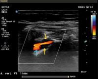 Fig. 6 Dissection of the vertebral artery. The distal segment of the extracranial vertebral
artery (V3) can be seen in the image. Typical of a dissection, a hypoechoic mural
hematoma (yellow arrows) can be seen stenosing the vessel, while a small aneurysm
has formed further along (blue arrow).
Fig. 6 Dissection of the vertebral artery. The distal segment of the extracranial vertebral
artery (V3) can be seen in the image. Typical of a dissection, a hypoechoic mural
hematoma (yellow arrows) can be seen stenosing the vessel, while a small aneurysm
has formed further along (blue arrow).
VA dissections most commonly occur in the V3 segment above or below the first cervical
vertebra or when passing through the dura in the foramen magnum, as the vessel is
partially fixed there by connective tissue and can be injured during jerky shearing
movements. From here, the dissection may continue cranially to intracranially and
caudally over a longer distance. In vertebral artery dissections, a mural hematoma
can often be detected in the vascular segments between the transverse processes of
the cervical vertebral bodies. A double lumen, on the other hand, is rare. In the
case of a high-grade luminal narrowing at the level of the atlas loop, a “sloshing
phenomenon” can be detected in the V2 segment [30]. Hematoma-related vascular narrowing must not be confused with large vessel vasculitis,
which is often concentric and longer in distance ([Fig. 7]).
 Fig. 7 Differential diagnosis of concentric vasoconstriction. Halo phenomenon of the temporal
artery in a 75-year-old female patient with cranial arteritis (left, top); the vertebral
artery is also involved (left, bottom) and shows a marked hypoechogenic rim. Right:
B-mode image of the CCA of a young patient with Takayasu’s arteritis shows long-stretch
concentric involvement of the vessel wall.
Fig. 7 Differential diagnosis of concentric vasoconstriction. Halo phenomenon of the temporal
artery in a 75-year-old female patient with cranial arteritis (left, top); the vertebral
artery is also involved (left, bottom) and shows a marked hypoechogenic rim. Right:
B-mode image of the CCA of a young patient with Takayasu’s arteritis shows long-stretch
concentric involvement of the vessel wall.
In the presence of high-grade luminal narrowing at the level of the skull base, flow
obstruction can be detected by indirect criteria of increased pulsatility of the Doppler
spectral waveform of the ICA and CCA.
Stents in the internal carotid artery
Stents in the internal carotid artery
Due to a decrease in vessel compliance and change in measurable flow phenomena, flow
velocities are somewhat higher in stenosis within a stent than in “normal” constrictions.
A PSV > 225 cm/s for 50 % and a PSV > 350 cm/s for 70 % can be considered as a threshold
for in-stent residual stenosis [31].
-
The length of stenosis influences peak systolic flow velocities (higher for very short
and lower for long stenoses)
-
Tandem stenoses (additional stenosis of the intracranial ICA) result in lower peak
systolic flow velocities in the extracranial stenosis maximum
-
An arteriovenous fistula fed by the ECA leads to “internalization” of the Doppler
spectrum of the ECA with high end-diastolic flow velocities
-
Calcified plaques with acoustic shadowing may make flow measurement impossible, and
confusion with occlusions is possible.
-
Large vessel vasculitis (giant cell arteritis, Takayasu’s arteritis) results in concentric,
homogeneously hypoechoic wall thickening: Confusion with dissections is possible
-
Abnormal origin of the ascending pharyngeal artery arising from and running parallel
to the ICA (may rejoin the ICA as a collateral and bridge a short ICA occlusion)
-
VA fenestration (normal Doppler spectrum in both lumen): Confusion with dissection
(pathological Doppler spectrum in at least one of the two lumen)
-
A. lusoria (right subclavian artery arises from the descending aorta instead of the
brachiocephalic trunk)
-
Truncus bicaroticus (both carotids arise from a common truncus)
-
All findings should be understandable based on the image and curve documentation
-
The multiparametric approach to ICA stenosis grading is based on both peak systolic
and diastolic flow velocities and B-scan morphologic criteria; in high-grade stenoses,
it is additionally based on indirect criteria and the presence of collateral circulation.
-
A multiparametric approach enables grading of high-grade ICA stenosis in 10 % increments
-
Intima-media thickness (IMT) is determined in a plaque-free region 2 cm proximal to
the bulb in the CCA on the posterior vessel wall
-
In B-mode imaging, atherosclerotic plaques are described using echogenicity, internal
structure, and surface
-
The origin of the vertebral artery from the subclavian artery is a predisposition
site for stenosis
-
Criteria for stenosis of the vertebral artery at the origin from the subclavian artery
is an angle-corrected PSV > 120 cm/s
-
There is hypoplasia of the VA with an absolute lumen diameter ≤ 2.0–2.5 mm in several
segments or a diameter ratio compared to the opposite side > 1:1.7
-
Higher-grade stenosis of the subclavian artery leads to a subclavian steal syndrom
of the ipsilateral vertebral artery
-
Criteria of a subclavian steal syndrome of the VA include systolic deceleration (grade
1) of the flow profile, alternating flow (grade 2), or completely retrograde flow
(grade 3) in the VA


