Keywords
characterization - chitosan - collagen - membrane - guided tissue regeneration
Introduction
The ultimate goal of periodontal therapy is to restore periodontal attachment, including cementum, periodontal ligament, and alveolar bone loss caused by periodontal disease.[1] Tissue engineering approaches have been introduced as an innovative alternative to conventional biomaterials to replace the lost or damaged human oral tissues. Various studies have attempted to regenerate dental tissues using a variety of scaffold materials such as natural polymers ( collagen, polysaccharides, or fibrin).[2] In dentistry, collagen is used as a membrane for guided tissue regeneration (GTR) or guided bone regeneration (GBR).[3] Ideal requirements for GTR membranes include occlusive cells that meet minimum mechanical, physical, structural, and biocompatibility requirements; ability to support vascularization and wound stabilization; protect blood clots thereby limiting epithelial connective tissue growth and unwanted into the defect; promote regeneration of functional tissue from cells that are relevant in the defect (avoid healing by repair); and provide space for newly formed periodontal tissues.[4]
[5]
Collagen is the main structural component of connective tissue that covers nearly 30% of the total protein in the body system. Collagen has been widely used as an essential compound in the food industry, pharmaceuticals, cosmetics, and biomedicals.[6] GTR or GBR membranes generally originate from collagen derived from the bone raw materials and mammalian skin, such as cattle and pigs. Raw materials from pigs, however, are not justified for Muslims and Jews, while the use of cow bones and skin is restricted for Hindus and raises concerns due to the presence of bovine spongiform encephalopathy or is also known as mad cow disease.[7] Consumption of bovine collagen allows transmission of infectious diseases toward humans. Therefore, an alternative collagen sourced from fish is currently being developed.
Waste from fisheries processing is known to reach up to 75% of the total of fish’s weight. The wastes are in the form of bones, skin, and fish scales, which are considered as lowvalue materials.[8] The processing of fisheries waste is expected to be able to reduce the waste volume while providing added value to the products manufactured.[9] Collagen is one of the materials contained in prospective fisheries waste to be developed. More than 30% of fishery waste as mentioned above is very rich in collagen, which can become a better alternative source of collagen. Various studies have reported extraction of collagen from Nile perch fish (Lates niloticus) skin,[10] big eye snapper (Priacanthus tayenus),[8] and grass carp (Ctenopharyngodon idella).[11] In addition to fish skin, fish scales also can become a collagen source. In this study, the source of collagen was extracted from white snapper scales. Barramundi is one type of edible portion fish in Indonesia whose wastes can be used as the collagen source. Indonesia as a maritime country has a great potential to develop fish collagen. Collagen from barramundi scales has a more stable collagen structure and is difficult to be degraded, with a porous surface, and is a quite good tensile strength.[12]
[13]
Other natural ingredients that are often used in the medical field are chitosan. Chitosan is an amino polysaccharide, which has attracted significant scientific interest during the past few decades. It is a partially deacetylated form of chitin, well known for showing biocompatibility, biodegradability, and antimicrobial activity.[14]
[15] Chitosan has been processed in various forms used in tissue engineering applications, such as membranes, nanoparticles, gels, scaffold, and sponge structure[16]
[17]. Previous research of barramundi collagen is used as wound dressing and corneal tissue engineering. To the best of our knowledge, research on membranes sourced from fish collagen is still being conducted and the research is still limited. The purpose of this study was to evaluate the synthesis, mechanical strength, and morphology of chitosan-collagen membranes from barramundi scales for GTR technique.
Materials and Methods
Materials and tools used for the fabrication of collagen membranes were as follows: the barramundi scales, chitosan (Surindo Co., Jakarta, Indonesia), 100% acetic acid, potassium bromide, distilled water, ethanol, sodium hydroxide, phosphate buffer saline, homogeneous, petri dish, Fourier transform infrared (FTIR), universal testing machine, and scanning electron microscope (SEM).
Collagen Extraction from Barramundi Scales
Collagen extraction was performed by immersing the barramundi scales in the acetic acid, as referred to the study of Muyonga et al in 2004. The collagen was extracted using acetic acid solution with a concentration of 0.5 M at 4°C for 3 days followed by regular stirring. Extraction was then repeated with the same reagents and conditions and then the obtained filtrate was centrifuged at 10,000 rpm for 30 minutes. Collagen precipitation was performed by adding NaCl to the supernatant with the concentration of 0.9 M and then centrifuged again. Collagen purification was performed by reimmersion in the acetic acid solution and then precipitated again with the procedures similar as before. The wet collagen obtained was then dried with a freeze drier to obtain dry collagen.
Chitosan Membrane Fabrication from Barramundi Scales
Preparation of chitosan solution was performed by immersing the chitosan with a concentration of 2% in 1% acetic acid. As much as 2 g of chitosan was immersed in 100 mL of acetic acid so that the chitosan produced would be in a gel form. Addition of collagen was performed by adding as much as 1 g of collagen in the chitosan solution and then placed in the petri dish according to the required thickness. Afterward, the solution mixture was being frozen with the freeze drier, so that the frozen molecular water molecules would be removed or drawn to form a gas. This process was called sublimation. This sublimation process will cause membranes fabricated having pores. The membranes were cut to a certain size and packaged in a plastic wrap ([Fig. 1]), then sterilization process was performed using the gamma-ray irradiation at a dose of 25 kGy for 1 to 3 hours, depends on the amount of sterilized material.
 Fig. 1 Collagen membrane sized 1 × 1 cm, wrapped with sealed and sterilized plastic.
Chitosan-Collagen Membrane Characterization
FTIR Analyses
FTIR is a tool for measuring the radiation absorption of infrared regions at various wavelengths.[18] This test is used to identify functional groups or collagen-chitosan compounds. The membrane sample was placed on a disk printer and then vacuumed to remove the gas content. The printed disk was then entered to the FTIR tool, and measured at the wavelength of 400 to 4,000 cm–1.
Scanning Electron Microscopy
All samples were placed in a vacuum—the device used was the FEI Quanta FEG 650 SEM with 2,000 kV accelerating voltage. The photos taken were the surface and the cross sections of the membrane with magnifications of 100× and 500×.
Tensile Strength
Determination of the mechanical properties was performed by cutting the membrane with a size of 40 × 10 mm. The cutting ends were connected to the test equipment, and the tensile load was calculated with N as the load unit. The membrane was then being pulled at a certain speed until it breaks. The amount of the membrane tensile load at the time of breaking was recorded. This test was performed with a universal testing machine.
Results
Collagen extraction from barramundi scales using the acetic acid was referred to the research conducted by Moyunga et al.[10] The collagen membrane was fabricated from collagen derived from barramundi scales, which were combined with chitosan. The fabrication was performed at The National Nuclear Energy Agency of Indonesia, Jakarta office. Collagen from barramundi scales was fabricated into membranes by cutting into 1 × 1 cm pieces and sterilized using radiation isotopes. The resulting membrane was then tested for various mechanical properties that were tensile strength and elongation at break, membrane porosity, membrane sterilization, and SEM.
The FTIR analysis result showed the typical peak of a chitosan- collagen membrane ([Fig. 2]) was at the absorption wavenumber of 3,453.9 cm–1 as a hydroxyl (–OH) group and also as an NH2 group, while at the peak of absorption wavenumber of 2,900 cm–1 is C–H, on the absorption wavenumber of 1,581.9 cm–1 was–NH2 bending. The presence of CH–NH2 group was shown in the absorption wavenumber of 1,150.3 cm–1. On the absorption wavenumber band of 1050 cm–1, the–C–O–C–glycosidic chain was presented between the chitosan monomer. Collagen had the typical peak on the absorption wavenumber of 3,453.9 cm–1 that was the group of hydroxyl (–OH) and an amide group. On the absorption wavenumber of 2,900 cm–1, the C–H group was presented, and an amide group was presented at the absorption wavenumber of 1581.9 cm–1 ([Table 1]).
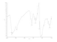 Fig. 2 Fourier transform infrared spectrum of chitosan-collagen membrane.
Table 1
FTIR data of chitosan-collagen membrane
|
Wavenumber (cm−1)
|
Chitosan
|
Collagen
|
|
Abbreviation: FTIR, Fourier transform infrared.
|
|
3453.9
|
–NH2
|
–OH
|
|
–OH
|
Amide
|
|
2900
|
C–H
|
C–H
|
|
1581.9
|
NH2 Bending
|
Amide
|
|
Primary alcohol
|
|
1150.3
|
CH–NH2
|
|
|
1050
|
–C–O–C glycosidic
|
|
Tensile strength is the maximum stress that a material can resist when stretched or pulled before the material breaks. Tensile strength and elongation (elasticity) of the membrane were measured in dry and wet conditions ([Table 2]). The tensile strength of the membrane was 0.28 MPa at the dry condition, and elongation at break was 8.53%, while in the wet condition, the tensile strength was 0.12 MPa and elongation at break was 25.6%.
Table 2
The membrane tensile strength and elongation at break
|
Membrane condition
|
Tensile strength
(MPa)
|
Elongation at break (%)
|
|
Dry
|
0.28
|
8.53
|
|
Wet
|
0.12
|
25.6
|
Porosity test is a measurement of spaces between the materials, and is a fraction of the volume of spaces to the total volume, expressed as the percentage between 0 and 100%. The membrane porosity test result in this study was 38.85%, which indicated that the spaces formed on the membrane were ~38.85% of the total volume of the membrane.
In this study, membrane sterilization was performed using the 25 kGy gamma irradiation rays, which resulted that until 14 days, there was no microbial growth in the thioglycolate medium ([Table 3]). Sterilization at a dose of 25 kGy was quite effective for sterilizing the membrane from microbial growth.
Table 3
Membrane sterility test
|
Microbes growth (days)
(thioglycollate medium)
|
|
1
|
2
|
3
|
4
|
5
|
6
|
7
|
14
|
|
Control
|
+
|
+
|
+
|
+
|
+
|
+
|
+
|
+
|
|
Tested membrane
|
−
|
−
|
−
|
−
|
−
|
−
|
−
|
−
|
SEM is an electron microscope designed to observe the surface of a solid object directly. SEM focuses on an electron beam on the object’s surface and captures its image by detecting electron appearance from the object surface. The scanning result of the collagen membrane using the SEM showed that the collagen elements were able to fuse with other elements, which were the chitosan and the mixture was homogeneous. The membrane surface was porous with sizes varying around 16 to 100 µm ([Fig. 3]).
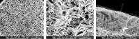 Fig. 3 Scanning electron microscope (SEM) result of collagen membrane with magnification 100x (A) and 500x (B); well-observed the homogeneity and porosity of the membrane surface. (C) SEM result of cross-sectional cut of collagen membrane with magnification of 500x, well-observed the fibrous and interconnected porous of membrane surface.
Discussion
The results of this study concluded that the chitosan- collagen membrane from barramundi scale showed the fibrous membrane surface, interconnected porous and ideal porous size as GTR membrane, and the lower mechanical strength. The previous research of collagen barramundi scale was done by Krishnan et al[19] and Sankar et al.[12] Krishnan et al used a collagen of barramundi scale as a corneal tissue engineering material; this study concluded that collagen from barramundi scales has good mechanical and physicochemical characterization, and potential biomedical materials that can be used in treatment.[19] Sankar et al stated that collagen from barramundi scales has good physical characteristics to be used as wound dressing material which has porous and fibrous in morphology as shown in SEM images.[12]
The success of an integration of two polymer components depends on the intermolecular interactions, which will result in an improved quality of the alloy mechanical properties. Tensile strength and elongation at break indicate the strength and elasticity of the membrane that are an important physical parameter in supporting the clinical application of the membrane.[3] Based on the results of the tensile strength measurement as presented in [Table 2], the fabricated membrane tensile strength in dry condition was 0.28 and 0.12 MPa in a wet condition. The membrane tensile strength was less than 1 MPa because the main element of this membrane was purely natural materials with no use of other synthetic chemicals to improve its mechanical properties. When compared with some existing membrane in the market, the tensile strength is still lower. Bio-Gide (Geistlich Biomaterials, Baden-Baden, Germany) membrane shows that maximum tensile stress and maximum tensile strain are 4.8 MPa and 46.8%, respectively, while Collprotect (Botiss Biomaterial, Italy) membrane shows that maximum tensile stress and maximum tensile strain are 13.1 MPa and 13.1%, respectively.[20]
The mechanical strength and chemical stability of the collagen membrane can be increased chemically by the use of compounds such as aldehyde and glutaraldehyde, and physically by ultraviolet and gamma irradiation.[3] The use of gamma irradiation is intended not only to modify the membrane physical properties but also to sterilize the membrane.[21] Our study was using the 25 kGy gamma irradiation to sterilize the collagen membrane. Gamma-ray irradiation can damage hydrogen bonds between those two macromolecules; the high doses are used to cause degradation of chitosan chains. Application of gamma irradiation on a mixture of chitosan/collagen membranes can reduce tensile strength and extend breaking.[22]
Gamma radiation and electron beam are the most popular tools used for material sterilization in the health sector and food preservation. Both of these tools can be used to coat the material surface. The advantage of gamma radiation is the relatively fast process, no residue, no catalyst, or no initiator needed, and irradiation doses can be adjusted as required.[22] In our study, gamma radiation is used to sterilize the fabricated membrane at a dose of 25 kGy. The membrane sterility test result is presented in [Table 3] and showed negative microbial growth until day 14, in other words the use of gamma radiation was effective to sterilize the membrane.
The physical characteristics of the scaffold are an essential factor in tissue engineering. The three-dimensional scaffold structure becomes an extracellular matrix that is similar or analogous as a template and physical support to guide cell proliferation and differentiation.[23] The three-dimensional scaffold must have high porosity and porous interconnected structures that will be able to facilitate cell attachment, proliferation, and differentiation.[24]
[25] Based on the SEM analysis as shown in [Fig. 3], the elements of collagen and chitosan were mixed homogeneously, and the porous size of the membrane surface was approximately 16 to 100 µm. The porous size in our study was similar to the previous study conducted by Mighri et al[26] and Rajam et al.[28] Research by Mighri et al showed that the mixture of collagen and chitosan was able to show a homogeneous surface in the porous size and distribution. The study also stated that the size and porous distribution of the nonchitosan-coated collagen membrane and the chitosan-coated collagen membrane showed a varied surface.[26] The porous size of the nonchitosan-coated membrane was higher (30–100 µm) compared with the chitosan-coated membrane (30–70 µm).[27] The result of another study conducted by Rajam et al showed that the membrane or scaffold of collagen and chitosan had interconnected porous with an average diameter of 75 to 150 µm.[28] The membranes showed a porous microarchitecture and some possibility of interconnectivity providing space for vascularization ([Fig. 3]). It has been reported that the minimum recommended pore size for a scaffold is 100 µm to achieve adequate vascularization of the tissue/organ being repaired or regenerated.[5]
[29]
Porous scaffold structure is influenced by the freezedrying method. During the freezing process, ice grain will be formed. The process of removing the crystals is performed by lyophilization so that the porous membrane will be created.[22]
[24] Membrane surface with interconnected porous will be able to facilitate cell attachment, proliferation, and differentiation that play essential roles in the wound healing and tissue regeneration. Natural ingredients such as collagen and chitosan are more widely used in clinical applications because of their roles in facilitating cell attachment and growth, and also tissue regeneration.[26]
Management and treatment of bone defects are a major clinical problem in the field of periodontology and oral implantology.[30] The final goal of periodontal therapy is to eliminate the inflammation and achieve periodontal tissue regeneration.[31] The mechanisms of GTR/GBR are to isolate periodontal bone defects from gingival connective tissue so that the new bone growth will be formed along with the alveolar bone defect.[32]
[33] Barrier membrane with a favorable function has to meet certain important design criterion that is biocompatibility. It should not stimulate the immune system or produce sensitization that may interfere with the wound healing process. To inhibit the migration of unwanted cells toward the material, the membrane should act as a barrier. This barrier, however, would allow the passage of nutrients and gases. Another important property of barrier material is tissue integration. This property inhibits a rapid epithelial migration on the material outer surface or material encapsulation. Ability to create and maintain a space adjacent to the root surface is another essential property. This property allows the cells from the periodontal ligament to entering the space. The membrane should have a design that is easy to trim and adjust to the desired site. Easy manipulation may affect the predictability of the clinical outcome.[34]
Conclusion
Within the limitations of this study, it can be concluded that chitosan-collagen membranes showed a fibrous membrane surface, ideal porous size recommended as GTR membrane; although it shows lower mechanical strength, this membrane has the potential to develop as an alternative membrane barrier for GTR. Further research is awaited to improve the mechanical strength of chitosan-collagen membranes.


