
Subscribe to RSS
DOI: 10.1055/s-0040-1721844
Surgical Treatment of Multiple Osteoporotic Fractures of the Dorsolumbar Spine: Case Report[*]
Article in several languages: português | English
Abstract
Osteoporotic vertebral fractures are a common type of fracture and affect a significant number of subjects with osteoporosis. Despite the high fracture risk, the concomitant occurrence of vertebral fractures at non-contiguous levels is very rare. We report the case of a patient with three burst dorsolumbar spine fractures at non-contiguous levels who was treated with percutaneous kyphoplasty and transpedicular posterior fixation. Six months after the surgery, the patient walks autonomously and without pain; in addition, there is no radiological evidence of fracture reduction loss.
#
Introduction
Compression vertebral fractures classified as AO Spine type A are among the most relevant clinical consequences from osteoporosis, and their incidence in the older population is increasing.[1] They are potential causes for chronic pain, leading to functional limitation and having a significant impact on physical activities, quality of life, and mortality. Fractures can be spontaneous or due to a traumatic event, usually resulting from a compressive mechanism involving the vertebral body. These fractures often involve the middle third of the thoracic spine and the dorsolumbar region.
Although most of these injuries are stable and feasible to conservative treatment based on rest, pain control and immobilizing orthoses, a considerable number of fractures, particularly in the dorsolumbar transition, can result in progressive height loss and increase kyphosis deformity; others may cause posterior wall rupture, leading to neural compression and neurological deficit. As such, early surgical stabilization is warranted. However, published studies show high rates of complications in osteoporotic patients undergoing surgical procedures such as kyphoplasty and posterior instrumentation, with selected series reporting adjacent vertebral fractures and material failure in about one third of these patients.[2]
#
Case Description
We report the case of a very active 70-year-old woman with osteoporosis who suffered a fall from her own height at home following a syncope. She immediately presented pain at the dorsolumbar transition. After 3 days of intense, persistent pain, despite anti-inflammatory and analgesic medication, she went to the hospital. She had pain on palpation of the spinous apophysis of the dorsolumbar transition, no muscle strength deficit in the lower limbs and normal osteotendinous reflexes. Fractures of the D9, D12, and L2 vertebral bodies, classified as type A4 according to the AO Spine system, were diagnosed in an osteoporotic context ([Figure 1]) in a subject with no associated conditions or chronically treated with osteoporosis-predisposing medications. The fractures showed signs of significant crushing of the intrabody bone trabeculae.

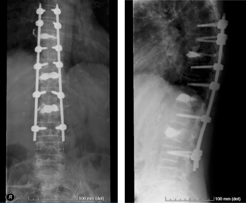
The patient underwent surgical treatment 4 days after hospital admission ([Figure 2]). Kyphoplasty was performed at the D9, D12, and L2 levels using intrabody stents through a transpedicular percutaneous approach with high viscosity polymethylmethacrylate (PMMA) filling. Since the D9 vertebral body could not bear two stents, even in their smallest size (small), this level received a single stent and cement filling. A percutaneous transpedicular posterior D7-D8-D10-D11-L1-L3 vertebrae fixation was performed using bars molded according to the patient's anatomy in thoracic kyphosis and lumbar lordosis (VIPER system, DePuy/Synthes, Warsaw, IN, USA) ([Figure 3]).


The patient could stand up on the first postoperative day and then walked with a dorsolumbar support. Six weeks after the surgery, she could walk alone presenting only residual pain. Anti-osteoporotic treatment started with denosumab, and, 6 months after surgery, the patient walks alone, with no pain complaints or loss of fracture reduction, and with favorable healing ([Figure 4]).
#
Discussion
The patient described in the present article presents a rare case of osteoporotic fractures of several non-contiguous levels of the dorsolumbar spine submitted to percutaneous treatment, assuring not only anterior support by kyphoplasty but also posterior tension band support through posterior fixation. This choice is justified by the high risk of progressive kyphosis development. Nevertheless, the instrumentation is considerably extent, and extraction may be considered after fractures consolidation.
The main risks in osteoporotic vertebral fractures include the progressive collapse of the vertebral body up to vertebra plana formation and gradual development of kyphotic deformities.[3] In fact, this sagittal balance disturbance is critical in elderly patients with osteoporosis due to the higher probability of progressive worsening of spinal sagittal changes. As a result, the paravertebral muscle tension increases, thus causing chronic pain, and sagittal imbalance may even lead to new fractures. In addition, a higher number of vertebral fractures will further anteriorly displace the spine's center of gravity.
Kyphoplasty with polymethylmetacrylate (PMMA) application is a well-documented procedure to correct and prevent collapse and deformities, with an important role in restoring harmony and overall sagittal balance of the spine.[4] However, in 10 to 30% of cases, the correction obtained by this type of procedure alone can gradually fail and increase kyphosis.[5] On the other hand, up to 25% of these cases develop fractures at an adjacent level, often the upper osteoporotic vertebra.[3]
In our case, the presence of three concomitant burst fractures in D9, D12, and L2, with a high risk of posttraumatic collapse, increased considerably the risk of complications of the posterior fixation alone. The strength of each posterior instrumentation fixation point is lower at the osteoporotic spine since the pullout force, the cut-out torque and the maximum insertion torque are directly proportional to the bone mineral density.[6] In patients undergoing spinal surgical treatment, osteoporosis has been associated with postinstrumentation vertebral fractures, pseudarthrosis, and secondary material failure.[7] Biomechanical studies have shown that insufficient anterior column support, along with the poor fixation provided by bones with low mineral density, may account for these unsuccessful outcomes.[8] Furthermore, the cavity formed at the fractured vertebral body after distraction delays consolidation and favors reduction loss.[9] Thus, we decided to combine a kyphoplasty with stent and percutaneous posterior fixation. Stents were selected because they provide greater vertebral body rigidity and reduce the risk of cement overflow, since they create an intrabody cavity surrounded by the implant. In compressive fractures, this hybrid technique was associated with less pain during the immediate postoperative period and spared virtually the whole height of the fractured vertebral body, with a low risk of recurrent kyphosis.[10] Therefore, we believe that it mitigates the risk of anterior collapse due to the lack of anterior column support at the posterior fixation, as well as the risk of fixation material failure and adjacent osteoporotic fractures.
#
#
Study developed at the Orthopedics and Traumatology Department, Centro Hospitalar e Universitário de Coimbra, Portugal.
-
Referências
- 1 Johnell O, Kanis JA. An estimate of the worldwide prevalence and disability associated with osteoporotic fractures. Osteoporos Int 2006; 17 (12) 1726-1733
- 2 Goldstein CL, Brodke DS, Choma TJ. Surgical Management of Spinal Conditions in the Elderly Osteoporotic Spine. Neurosurgery 2015; 77 (Suppl. 04) S98-S107
- 3 Frankel BM, Monroe T, Wang C. Percutaneous vertebral augmentation: an elevation in adjacent-level fracture risk in kyphoplasty as compared with vertebroplasty. Spine J 2007; 7 (05) 575-582
- 4 Papanastassiou ID, Phillips FM, Van Meirhaeghe J. et al. Comparing effects of kyphoplasty, vertebroplasty, and non-surgical management in a systematic review of randomized and non-randomized controlled studies. Eur Spine J 2012; 21 (09) 1826-1843
- 5 Komemushi A, Tanigawa N, Kariya S. et al. Percutaneous vertebroplasty for osteoporotic compression fracture: multivariate study of predictors of new vertebral body fracture. Cardiovasc Intervent Radiol 2006; 29 (04) 580-585
- 6 Paxinos O, Tsitsopoulos PP, Zindrick MR. et al. Evaluation of pullout strength and failure mechanism of posterior instrumentation in normal and osteopenic thoracic vertebrae. J Neurosurg Spine 2010; 13 (04) 469-476
- 7 DeWald CJ, Stanley T. Instrumentation-related complications of multilevel fusions for adult spinal deformity patients over age 65: surgical considerations and treatment options in patients with poor bone quality. Spine (Phila Pa 1976) 2006; 31 (19 suppl): S144-S151
- 8 Norton RP, Milne EL, Kaimrajh DN, Eismont FJ, Latta LL, Williams SK. Biomechanical analysis of four- versus six-screw constructs for short-segment pedicle screw and rod instrumentation of unstable thoracolumbar fractures. Spine J 2014; 14 (08) 1734-1739
- 9 Knop C, Fabian HF, Bastian L, Blauth M. Late results of thoracolumbar fractures after posterior instrumentation and transpedicular bone grafting. Spine 2001; 26 (01) 88-99
- 10 Zhang J, Liu H, Liu H. et al. Intermediate screws or kyphoplasty: Which method of posterior short-segment fixation is better for treating single-level thoracolumbar burst fractures?. Eur Spine J 2019; 28 (03) 502-510
Endereço para correspondência
Publication History
Received: 25 May 2020
Accepted: 17 September 2020
Article published online:
22 March 2021
© 2021. Sociedade Brasileira de Ortopedia e Traumatologia. This is an open access article published by Thieme under the terms of the Creative Commons Attribution-NonDerivative-NonCommercial License, permitting copying and reproduction so long as the original work is given appropriate credit. Contents may not be used for commecial purposes, or adapted, remixed, transformed or built upon. (https://creativecommons.org/licenses/by-nc-nd/4.0/)
Thieme Revinter Publicações Ltda.
Rua do Matoso 170, Rio de Janeiro, RJ, CEP 20270-135, Brazil
-
Referências
- 1 Johnell O, Kanis JA. An estimate of the worldwide prevalence and disability associated with osteoporotic fractures. Osteoporos Int 2006; 17 (12) 1726-1733
- 2 Goldstein CL, Brodke DS, Choma TJ. Surgical Management of Spinal Conditions in the Elderly Osteoporotic Spine. Neurosurgery 2015; 77 (Suppl. 04) S98-S107
- 3 Frankel BM, Monroe T, Wang C. Percutaneous vertebral augmentation: an elevation in adjacent-level fracture risk in kyphoplasty as compared with vertebroplasty. Spine J 2007; 7 (05) 575-582
- 4 Papanastassiou ID, Phillips FM, Van Meirhaeghe J. et al. Comparing effects of kyphoplasty, vertebroplasty, and non-surgical management in a systematic review of randomized and non-randomized controlled studies. Eur Spine J 2012; 21 (09) 1826-1843
- 5 Komemushi A, Tanigawa N, Kariya S. et al. Percutaneous vertebroplasty for osteoporotic compression fracture: multivariate study of predictors of new vertebral body fracture. Cardiovasc Intervent Radiol 2006; 29 (04) 580-585
- 6 Paxinos O, Tsitsopoulos PP, Zindrick MR. et al. Evaluation of pullout strength and failure mechanism of posterior instrumentation in normal and osteopenic thoracic vertebrae. J Neurosurg Spine 2010; 13 (04) 469-476
- 7 DeWald CJ, Stanley T. Instrumentation-related complications of multilevel fusions for adult spinal deformity patients over age 65: surgical considerations and treatment options in patients with poor bone quality. Spine (Phila Pa 1976) 2006; 31 (19 suppl): S144-S151
- 8 Norton RP, Milne EL, Kaimrajh DN, Eismont FJ, Latta LL, Williams SK. Biomechanical analysis of four- versus six-screw constructs for short-segment pedicle screw and rod instrumentation of unstable thoracolumbar fractures. Spine J 2014; 14 (08) 1734-1739
- 9 Knop C, Fabian HF, Bastian L, Blauth M. Late results of thoracolumbar fractures after posterior instrumentation and transpedicular bone grafting. Spine 2001; 26 (01) 88-99
- 10 Zhang J, Liu H, Liu H. et al. Intermediate screws or kyphoplasty: Which method of posterior short-segment fixation is better for treating single-level thoracolumbar burst fractures?. Eur Spine J 2019; 28 (03) 502-510


