

Subscribe to RSS
DOI: 10.1055/s-0042-1751009
Panoramic Indices: An Aid to Early Detection of Patients with Low Bone Mineral Density
Authors
Funding None.

Abstract
Objective An early identification of patients who are at an obvious increased risk of osteoporosis and subsequent high risk of pathological bone fractures is important to prevent morbidity and its subsequent impact on the quality of life of the affected patients. Dental professionals have a chance of identifying such cases in their very initial stages through routinely prescribed dental radiographs. The aim of this study was to assess the influence of gender and age on different parameters of alveolar bone loss using orthopantomograph (OPG) as an aid to identify patients with low bone mineral density (BMD).
Materials and Methods This study included eighty subjects in whom after taking OPGs, mandibular cortical index (MCI), mandibular cortical width (MCW), and panoramic mandibular index were assessed, while the results obtained were subjected to statistical analysis. One-way analysis of variance followed by Tukey's test was used to compare the means, while p-value less than 0.05 was considered statistically significant.
Results The findings of this study revealed a significant association between MCI and age for females, with C2 and C3 categories being more common with advancing age. Also, a significant difference could be seen in relation to gender (p-value = 0.0315) for MCW with a concomitant decrease in the values of MCW in females over 60 years of age.
Conclusion Panoramic radiographic measurements could provide valuable information and help in screening patients with low BMD.
Introduction
Continuing alveolar bone loss (ABL) from edentulous jaws is a common concern that poses great challenge in the prosthetic rehabilitation in such patients. This bone loss is maximum during the initial 6 months postextraction during the wound healing period, though the process of resorption continues throughout life, even after the extraction sockets have healed, at a much slower pace. According to Devlin and Ferguson,[1] alveolar bone turnover is in part determined by a plethora of factors, anatomic and physiological, including diet, age, sex, and hormonal status of the individual in addition to the significant local factors that may interact in conjunct with the systemic factors. Also, elderly people are on the most risk for developing osteoporosis and associated fractures and this is more so in case of females with a still higher predisposition for developing osteoporosis with advancing age. The American Dental Association recommends screening radiography as a necessary step for all edentulous patients because of high prevalence of root fragments and various other pathological findings.[2] Also, few studies have suggested the applicability of oral radiology as an important diagnostic adjunct in the early detection of various systemic diseases including osteoporosis. Osteoporosis is a multifactorial disease process for which one of the major risk factors is the declining bone mass in postmenopausal females due to decreasing estrogen levels that increase their risk for fractures, if left undiagnosed or, untreated.[3] [4] [5] [6] Hence, an early identification of such cases in which there is an obvious risk for pathological fractures is all the more important to prevent morbidity and its subsequent impact on the quality of life (QoL) of the affected patients. Dental professionals have a chance of identifying this subclinical condition in its nascent stages through routinely prescribed dental radiographs wherein distinguished radiographic indices including mandibular cortical index (MCI), mandibular cortical width (MCW), and panoramic mandibular index (PMI) may be beneficial for identifying patients with low bone mineral density (BMD). Devlin et al[7] concluded MCW to be a more sensitive indicator than MCI in diagnosing such patients in their study while in another study, Benson et al[8] stated PMI to be a more sensitive indicator of low BMD. This study was planned with a similar intent to assess the influence of gender and age on different parameters of ABL using orthopantomograph (OPG) as an aid to identify patients with low BMD.
Materials and Methods
This observational study included 80 subjects (40 dentate and 60 completely edentulous) fulfilling the inclusion and exclusion criteria over a period of 1 year. The subjects were divided into two groups: dentate and edentulous groups. Dentate group comprised of 20 male and 20 female subjects in an age range of 41 to 75 years based on select inclusion criteria with the basic inclusion criteria of full complement of teeth with at least 28 teeth with or, without third molars in Angle's Class I occlusion. The selected edentulous subjects were divided into four groups (15 males and 15 females < 60 years and a similar number of subjects > 60 years of age) who were completely edentulous for more than 2 years. A written informed consent was obtained from all the participants before their inclusion into the study, while ethical clearance was obtained from the Institutional Ethics Committee via letter approval no. SDDC/IEC/08–41–2019. From dentate jaws, the location of the first premolar and molar regions was recorded and the approximate positions of the same were marked on radiographs of the edentulous subjects. Subjects with any kind of physical abnormality or, facial asymmetry, subjects who were edentulous for less than 2 years (since alveolar bone loss is more pronounced during the first few years after tooth loss), and subjects who were on long-term steroid therapy were excluded from this study apart from pregnant and lactating females to avoid risk of their exposure to radiation. All the subjects were subjected to general physical examination followed by a detailed and comprehensive oral examination under strict aseptic protocols. The subjects were, then, subjected for radiographic examination wherein all panoramic radiographs were taken with the same digital panoramic apparatus (Villa India Rotograph EVO) in Dental Studio Plus Window mode at 72 kVp and 6 mA exposure for 14.4 seconds to standardize the radiographic quality and to minimize errors related to technical detailing of the digital panoramic apparatus. Radiographic images with optimal diagnostic information and with no gross distortion of image were, then, selected to trace landmarks including the inferior and posterior border of mandible and mental foramen, first manually and then, with the help of an electronic digital caliper with accuracy up to 0.001 mm and the values obtained were subjected to statistical analysis.
Dentate Group
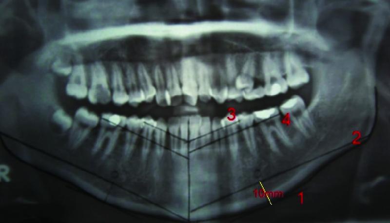
In the dentate group, on the digital radiograph obtained, a horizontal line was drawn tangential to the most inferior point of mandibular angle and lower border of the body of mandible and another line parallel to the above-mentioned tangential line and 10 mm above the inferior border of the mandible. The proportion of the horizontal mandibular length was determined by dividing the length of the mandibular body (from midline to the posterior border of ramus) by distance between the first premolar and midline. Thus, the locations of first premolar and molar were placed on the edentulous mandible ([Fig. 1]).
Edentulous Group: In the edentulous group, the following measurements were made:
MCI: It is the morphology of the mandibular inferior cortex as devised by Klemetti et al[9] who classified the morphology of mandibular inferior cortex as follows:
-
C1—The endosteal margin of the cortex is even and sharp on both sides of the body of the mandible;
-
C2—The endosteal margin has resorptive cavities with cortical residues, one to three layers thick, on one or, both sides of the body of the mandible; and
-
C3—The endosteal margin is clearly porous and consists of thick cortical residues.
MCW: It is the measurement of the cortical width at the region of mental foramen. For assessing the MCW, a line parallel to the long axis of mandible and tangential to inferior border of the mandible was drawn. A line perpendicular to this tangent intersecting the inferior border of mental foramen was, then, constructed and along this line, MCW was measured as the distance between the two parallel lines ([Fig. 2]).[10]

PMI: It is defined as the ratio of MCW to the distance between the superior or inferior margin of the mental foramen to the inferior border of the mandible, also, recognized as the basal bone height (BBH) ([Fig. 3]).[11]

ABL: It is calculated as the ratio of the radiographic total bone height to the BBH that is calculated from the center of the mental foramen to the inferior border of the mandible ([Fig. 3]).[12]
Statistical Analysis
Statistical analysis was performed using Epi Info (TM) 3.5.3. Descriptive statistical analysis was performed to calculate the means with corresponding standard deviations, while test of proportion was used to find standard normal deviate (Z) to compare the difference in proportions. Also, chi-squared test was used to find the associations, while one-way analysis of variance followed by Tukey's test was used to compare the means in more than two groups. Under Tukey's test, critical difference was calculated to compare pairwise means. p-Value less than 0.05 was considered statistically significant.
Results
Only two categories of MCI (C2 and C3) were detected in this study, while the association between MCI and age was not found to be statistically significant in males (p-value > 0.05), though significant in females (p-value = 0.0001). Also, females in the age group of less than 60 years were associated more with C2, while females more than 60 years were associated with C3 cortex. ([Table 1]) In terms of total mandibular length to distance at first premolar and first molar regions from midline in dentate males and females, the results were found to be significant only in relation to total mandibular length to distance at first molar region from midline (p-value = 0.0001) ([Table 2]). On further analysis, no significant difference was observed between age groups (p-value > 0.05), though a significant difference could be seen in relation to gender (p-value = 0.0315) for MCW. Furthermore, it was, also, observed that the mean values did not change with increase in age in males but decreased with increase in age in females; thus, a significant interaction could be observed between age and gender in terms of MCW ([Table 3]). Again, no significant difference was found between the medians of PMI (p-value > 0.05). For this parameter, there was no significant difference between age groups in this study as well as the values were not affected by age and/or gender (p-value > 0.05) ([Table 3]). Also, though the average bone height values were higher for males than females, no statistically significant difference was found between male and female subjects (p-value > 0.05). The bone loss was more in females of age group more than 60 years of age, though no significant change could be observed between the mean values of males and females of both the age groups making the study in favor of concluding that ABL was not affected by age and gender ([Table 3]).
|
Gender |
Age (in y) |
C2 (n) |
C3 (n) |
χ2-Value/p-Value |
|---|---|---|---|---|
|
Male |
≤ 60 |
14 |
1 |
χ2 = 1.1540; p = 0.2830 |
|
> 60 |
12 |
3 |
||
|
Female |
≤ 60 |
11 |
4 |
χ2 = 10.9950; p = 0.0001[a] |
|
> 60 |
2 |
13 |
a p < 0.05 is statistically significant.
|
Tooth |
Variable (in mm) |
Male |
Female |
t-Value |
p-Value |
|---|---|---|---|---|---|
|
Mean ± SD |
Mean ± SD |
||||
|
First premolar |
Mandibular length from midline |
85.63 ± 2.52 |
86.02 ± 1.60 |
−0.4132 |
0.6843 |
|
Distance at first premolar region from midline |
26.38 ± 0.93 |
26.96 ± 4.41 |
−0.4069 |
0.6889 |
|
|
First molar |
Mandibular length from midline |
85.63 ± 2.52 |
86.02 ± 1.60 |
−0.4132 |
0.6843 |
|
Distance at first molar region from midline |
43.97 ± 1.14 |
41.53 ± 0.73 |
5.6983 |
0.0001[a] |
Abbreviation: SD, standard deviation.
a p < 0.05 is statistically significant.
|
Gender and age (in y) |
MCW |
PMI |
ABL |
|
|---|---|---|---|---|
|
Mean ± SD |
Mean ± SD |
Mean ± SD |
||
|
Males ≤ 60 |
4.87 ± 1.03 |
0.37 ± 0.12 |
1.81 ± 0.34 |
|
|
Males > 60 |
4.87 ± 0.70 |
0.35 ± 0.12 |
2.04 ± 0.54 |
|
|
Females ≤ 60 |
4.62 ± 0.89 |
0.36 ± 0.12 |
1.85 ± 0.41 |
|
|
Females > 60 |
4.18 ± 0.61 |
0.35 ± 0.08 |
1.70 ± 0.47 |
|
|
Between gender |
F-Value |
4.8638 |
0.0005 |
1.6158 |
|
p-Value |
0.0315[a] |
0.9819 |
0.2089 |
|
|
Between ages |
F-Value |
1.0336 |
0.3249 |
0.1202 |
|
p-Value |
0.3137 |
0.5710 |
0.7301 |
|
Abbreviation: SD, standard deviation.
a p < 0.05 is statistically significant.
Discussion
Panoramic radiographs or OPGs have been the radiograph of choice for the initial screening of dental patients regarding any occult pathologic changes. In addition, the common drawbacks usually seen in OPGs in the form of unequal magnification and geometric distortion can be overcome by comparing proportions, rather than the actual measurements on such radiographs. In this context, panoramic radiographs or OPGs are still the most recommended radiographs in day-to-day practice for the gross screening of patients as well as to plan further radiographic investigations, in case, a need arises and/or, for treatment planning.[13] [14] [15] [16]
The results of this study suggested the association between MCI and age to be statistically insignificant in males, though significant in females (p-value = 0.0001), while only two categories of MCI (C2 and C3) could be observed in this study. Also, females in the age group of less than 60 years of age were found to be associated more with C2 than females of more than 60 years of age in whom C3 cortex was more common suggesting a possible shift to C3 cortex in aging females. The results of this study were in close accordance with the study conducted by Ledgerton et al[17] who found a close association between BMD detected by dual energy X-ray absorptiometry and the MCI scale (C1-C3) devised by Klemetti et al.[9] Bras et al,[18] also, concluded from the findings of their study that postmenopausal females and patients suffering with chronic renal failure had a comparatively thinner mandibular cortex than as compared with the controls. A notable observation made in this regard in the study conducted by Zlatarić and Celebić[19] suggested low BMD values in patients with C3 mandibular cortex, thus, concluding MCI to be a sensitive indicator of BMD.
Also, the findings of this study suggested no significant difference in terms of age (p-value > 0.05), though significant difference in relation to gender (p-value = 0.0315) for MCW. Furthermore, mean MCW was almost stable, while there was not much impact of age on the mean MCW in males; however, it was found to be lesser in females more than 60 years of age indicating a significant interaction between age and gender in terms of MCW in close accordance with the studies conducted by Ledgerton et al,[17] Dutra et al,[20] and Yüzügüllü et al.[21] Similar findings were observed in the study conducted by Zlatarić and Celebić [22] that observed a gradual decrease in the mean MCW up to 75 years of age in both males and females, though, after this age, the values were found to decrease sharply in females while being stable in males.
In terms of PMI, no significant differences could be observed in this study, while the values were not found to be affected by age and/or gender (p-value > 0.05). Ledgerton et al,[17] in their study, also, found a common pattern in PMI observing that up to sixth decade of life, there was seen a gradual decrease in PMI and thereafter, the values decreased sharply. Similar results were found in the studies conducted by Benson et al,[8] Ledgerton et al,[17] Yüzügüllü et al,[21] and Klemetti et al,[23] though higher PMI values were observed in the studies conducted by Benson et al,[8] Ledgerton et al,[17] and Klemetti et al[23] than the values observed in the study conducted by Yüzügüllü et al[21] that could be attributed to the ethnic variation in the populations studied. Also, Klemetti and Kolmakow[24] concluded from the results of their study that, though, a strong positive correlation could not be established between PMI and BMD, PMI, still could be used as an indicator of bone changes when there is large deviation from the mean PMI.
The results of this study, also, observed that, though the average bone height was found to be higher for males than females, the results were found to be statistically insignificant between the male and female subjects (p-value > 0.05) in respect to ABL. Furthermore, the findings of this study suggested significant bone loss in females of more than 60 years of age in close accordance with the studies conducted by Yüzügüllü et al[21] and Klemetti and Kolmakow.[24] The reason for more severe bone loss observed in females of age group more than 60 years of age could be attributed to the postmenopausal changes seen in this age group that are accompanied by a more generalized skeletal osteoporosis due to deficiency of hormone estrogen that accelerates skeletal bone loss with possibly, rapid alveolar bone resorption.
Conclusion
Within its own limitations and based on the findings obtained in this study, the study concludes that panoramic radiography might provide valuable information and could help in screening patients with low BMD who could, then, be referred for an appropriate and specialized care to prevent high-grade morbidity improving QoL of the affected individuals.
Conflict of Interest
None declared.
-
References
- 1 Devlin H, Ferguson MW. Alveolar ridge resorption and mandibular atrophy. A review of the role of local and systemic factors. Br Dent J 1991; 170 (03) 101-104
- 2 Alonso MB, Cortes AR, Camargo AJ, Arita ES, Haiter-Neto F, Watanabe PC. Assessment of panoramic radiomorphometric indices of the mandible in a Brazilian population. ISRN Rheumatol 2011; 2011: 854287
- 3 Nemati S, Kajan ZD, Saberi BV, Arzin Z, Erfani MH. Diagnostic value of panoramic indices to predict osteoporosis and osteopenia in post-menopausal women. J Oral Maxillofac Radiol 2016; 4: 23-30
- 4 Calciolari E, Donos N, Park JC, Petrie A, Mardas N. Panoramic measures for oral bone mass in detecting osteoporosis: a systematic review and meta-analysis. J Dent Res 2015; 94 (3, Suppl): 17S-27S
- 5 Mohammed AR, Abbas FK, Hassan NA. Diagnostic efficacy of mandibular cortical thickness on panoramic radiographs to identify post-menopausal women with low bone mineral densities (Iraqi population). J Am Sci 2013; 9: 308-312
- 6 Jagelavičienė E, Krasauskienė A, Žalinkevičius R, Vaitkevičienė I, Kubilius R. Relationship between the mandibular cortical index and calcaneal bone mineral density in postmenopausal women. Medicina (Kaunas) 2016; 52 (02) 125-131
- 7 Devlin H, Karayianni K, Mitsea A. et al. Diagnosing osteoporosis by using dental panoramic radiographs: the OSTEODENT project. Oral Surg Oral Med Oral Pathol Oral Radiol Endod 2007; 104 (06) 821-828
- 8 Benson BW, Prihoda TJ, Glass BJ. Variations in adult cortical bone mass as measured by a panoramic mandibular index. Oral Surg Oral Med Oral Pathol 1991; 71 (03) 349-356
- 9 Klemetti E, Kolmakov S, Kröger H. Pantomography in assessment of the osteoporosis risk group. Scand J Dent Res 1994; 102 (01) 68-72
- 10 Hekmatin E, Ahmadi SS, Ataiekhorasgani M, Feizianfard M, Jafaripozve S, Jafaripozve N. Prediction of lumbar spine bone mineral density from the mandibular cortical width in postmenopausal women. J Res Med Sci 2013; 18 (11) 951-955
- 11 Johari Khatoonabad M, Aghamohammadzade N, Taghilu H, Esmaeili F, Jabbari Khamnei H. Relationship among panoramic radiography findings, biochemical markers of bone turnover and hip BMD in the diagnosis of post-menopausal osteoporosis. Iran J Radiol 2011; 8 (01) 23-28
- 12 Ishii K, Taguchi A, Nakamoto T. et al. Diagnostic efficacy of alveolar bone loss of the mandible for identifying postmenopausal women with femoral osteoporosis. Dentomaxillofac Radiol 2007; 36 (01) 28-33
- 13 Gulsahi A, Yüzügüllü B, Imirzalioglu P, Genç Y. Assessment of panoramic radiomorphometric indices in Turkish patients of different age groups, gender and dental status. Dentomaxillofac Radiol 2008; 37 (05) 288-292
- 14 Govindraju P, Chandra P. Radiomorphometric indices of the mandible - an indicator of osteoporosis. J Clin Diagn Res 2014; 8 (03) 195-198
- 15 Cakur B, Dagistan S, Sahin A, Harorli A, Yilmaz A. Reliability of mandibular cortical index and mandibular bone mineral density in the detection of osteoporotic women. Dentomaxillofac Radiol 2009; 38 (05) 255-261
- 16 Gaur B, Chaudhary A, Wanjari PV, Sunil M, Basavaraj P. Evaluation of panoramic radiographs as a screening tool of osteoporosis in post-menopausal women: a cross-sectional study. J Clin Diagn Res 2013; 7 (09) 2051-2055
- 17 Ledgerton D, Horner K, Devlin H, Worthington H. Radiomorphometric indices of the mandible in a British female population. Dentomaxillofac Radiol 1999; 28 (03) 173-181
- 18 Bras J, van Ooij CP, Abraham-Inpijn L, Kusen GJ, Wilmink JM. Radiographic interpretation of the mandibular angular cortex: a diagnostic tool in metabolic bone loss. Part I. Normal state. Oral Surg Oral Med Oral Pathol 1982; 53 (05) 541-545
- 19 Zlatarić DK, Celebić A. Clinical bone densitometric evaluation of the mandible in removable denture wearers dependent on the morphology of the mandibular cortex. J Prosthet Dent 2003; 90 (01) 86-91
- 20 Dutra V, Yang J, Devlin H, Susin C. Mandibular bone remodelling in adults: evaluation of panoramic radiographs. Dentomaxillofac Radiol 2004; 33 (05) 323-328
- 21 Yüzügüllü B, Gulsahi A, Imirzalioglu P. Radiomorphometric indices and their relation to alveolar bone loss in completely edentulous Turkish patients: a retrospective study. J Prosthet Dent 2009; 101 (03) 160-165
- 22 Knezović-Ziatarić D, Celebić A. Mandibular bone mineral density changes in complete and removable partial denture wearers: a 6-month follow-up study. Int J Prosthodont 2003; 16 (06) 661-665
- 23 Klemetti E, Kolmakov S, Heiskanen P, Vainio P, Lassila V. Panoramic mandibular index and bone mineral densities in postmenopausal women. Oral Surg Oral Med Oral Pathol 1993; 75 (06) 774-779
- 24 Klemetti E, Kolmakow S. Morphology of the mandibular cortex on panoramic radiographs as an indicator of bone quality. Dentomaxillofac Radiol 1997; 26 (01) 22-25
Address for correspondence
Publication History
Article published online:
24 August 2022
© 2022. Asian Congress of Neurological Surgeons. This is an open access article published by Thieme under the terms of the Creative Commons Attribution-NonDerivative-NonCommercial License, permitting copying and reproduction so long as the original work is given appropriate credit. Contents may not be used for commercial purposes, or adapted, remixed, transformed or built upon. (https://creativecommons.org/licenses/by-nc-nd/4.0/)
Thieme Medical and Scientific Publishers Pvt. Ltd.
A-12, 2nd Floor, Sector 2, Noida-201301 UP, India
-
References
- 1 Devlin H, Ferguson MW. Alveolar ridge resorption and mandibular atrophy. A review of the role of local and systemic factors. Br Dent J 1991; 170 (03) 101-104
- 2 Alonso MB, Cortes AR, Camargo AJ, Arita ES, Haiter-Neto F, Watanabe PC. Assessment of panoramic radiomorphometric indices of the mandible in a Brazilian population. ISRN Rheumatol 2011; 2011: 854287
- 3 Nemati S, Kajan ZD, Saberi BV, Arzin Z, Erfani MH. Diagnostic value of panoramic indices to predict osteoporosis and osteopenia in post-menopausal women. J Oral Maxillofac Radiol 2016; 4: 23-30
- 4 Calciolari E, Donos N, Park JC, Petrie A, Mardas N. Panoramic measures for oral bone mass in detecting osteoporosis: a systematic review and meta-analysis. J Dent Res 2015; 94 (3, Suppl): 17S-27S
- 5 Mohammed AR, Abbas FK, Hassan NA. Diagnostic efficacy of mandibular cortical thickness on panoramic radiographs to identify post-menopausal women with low bone mineral densities (Iraqi population). J Am Sci 2013; 9: 308-312
- 6 Jagelavičienė E, Krasauskienė A, Žalinkevičius R, Vaitkevičienė I, Kubilius R. Relationship between the mandibular cortical index and calcaneal bone mineral density in postmenopausal women. Medicina (Kaunas) 2016; 52 (02) 125-131
- 7 Devlin H, Karayianni K, Mitsea A. et al. Diagnosing osteoporosis by using dental panoramic radiographs: the OSTEODENT project. Oral Surg Oral Med Oral Pathol Oral Radiol Endod 2007; 104 (06) 821-828
- 8 Benson BW, Prihoda TJ, Glass BJ. Variations in adult cortical bone mass as measured by a panoramic mandibular index. Oral Surg Oral Med Oral Pathol 1991; 71 (03) 349-356
- 9 Klemetti E, Kolmakov S, Kröger H. Pantomography in assessment of the osteoporosis risk group. Scand J Dent Res 1994; 102 (01) 68-72
- 10 Hekmatin E, Ahmadi SS, Ataiekhorasgani M, Feizianfard M, Jafaripozve S, Jafaripozve N. Prediction of lumbar spine bone mineral density from the mandibular cortical width in postmenopausal women. J Res Med Sci 2013; 18 (11) 951-955
- 11 Johari Khatoonabad M, Aghamohammadzade N, Taghilu H, Esmaeili F, Jabbari Khamnei H. Relationship among panoramic radiography findings, biochemical markers of bone turnover and hip BMD in the diagnosis of post-menopausal osteoporosis. Iran J Radiol 2011; 8 (01) 23-28
- 12 Ishii K, Taguchi A, Nakamoto T. et al. Diagnostic efficacy of alveolar bone loss of the mandible for identifying postmenopausal women with femoral osteoporosis. Dentomaxillofac Radiol 2007; 36 (01) 28-33
- 13 Gulsahi A, Yüzügüllü B, Imirzalioglu P, Genç Y. Assessment of panoramic radiomorphometric indices in Turkish patients of different age groups, gender and dental status. Dentomaxillofac Radiol 2008; 37 (05) 288-292
- 14 Govindraju P, Chandra P. Radiomorphometric indices of the mandible - an indicator of osteoporosis. J Clin Diagn Res 2014; 8 (03) 195-198
- 15 Cakur B, Dagistan S, Sahin A, Harorli A, Yilmaz A. Reliability of mandibular cortical index and mandibular bone mineral density in the detection of osteoporotic women. Dentomaxillofac Radiol 2009; 38 (05) 255-261
- 16 Gaur B, Chaudhary A, Wanjari PV, Sunil M, Basavaraj P. Evaluation of panoramic radiographs as a screening tool of osteoporosis in post-menopausal women: a cross-sectional study. J Clin Diagn Res 2013; 7 (09) 2051-2055
- 17 Ledgerton D, Horner K, Devlin H, Worthington H. Radiomorphometric indices of the mandible in a British female population. Dentomaxillofac Radiol 1999; 28 (03) 173-181
- 18 Bras J, van Ooij CP, Abraham-Inpijn L, Kusen GJ, Wilmink JM. Radiographic interpretation of the mandibular angular cortex: a diagnostic tool in metabolic bone loss. Part I. Normal state. Oral Surg Oral Med Oral Pathol 1982; 53 (05) 541-545
- 19 Zlatarić DK, Celebić A. Clinical bone densitometric evaluation of the mandible in removable denture wearers dependent on the morphology of the mandibular cortex. J Prosthet Dent 2003; 90 (01) 86-91
- 20 Dutra V, Yang J, Devlin H, Susin C. Mandibular bone remodelling in adults: evaluation of panoramic radiographs. Dentomaxillofac Radiol 2004; 33 (05) 323-328
- 21 Yüzügüllü B, Gulsahi A, Imirzalioglu P. Radiomorphometric indices and their relation to alveolar bone loss in completely edentulous Turkish patients: a retrospective study. J Prosthet Dent 2009; 101 (03) 160-165
- 22 Knezović-Ziatarić D, Celebić A. Mandibular bone mineral density changes in complete and removable partial denture wearers: a 6-month follow-up study. Int J Prosthodont 2003; 16 (06) 661-665
- 23 Klemetti E, Kolmakov S, Heiskanen P, Vainio P, Lassila V. Panoramic mandibular index and bone mineral densities in postmenopausal women. Oral Surg Oral Med Oral Pathol 1993; 75 (06) 774-779
- 24 Klemetti E, Kolmakow S. Morphology of the mandibular cortex on panoramic radiographs as an indicator of bone quality. Dentomaxillofac Radiol 1997; 26 (01) 22-25