

Subscribe to RSS
DOI: 10.1055/s-0044-1786981
Childhood Solid Pseudopapillary Neoplasm of the Pancreas: Acute Presentation of an Uncommon Tumor
Authors


Abstract
Solid pseudopapillary tumor is a mixed solid and cystic neoplasm of the pancreas which occurs most commonly in females in second or third decade of life with few reported pediatric cases. It has a low malignancy potential with good prognosis after surgical resection.
We present a case of an 11-year-old male child with solid pseudopapillary tumor of pancreas who presented with clinical features of acute pancreatitis. He was initially evaluated with ultrasound, followed by computed tomography (CT) and magnetic resonance imaging (MRI).
Solid pseudopapillary tumor of pancreas presenting as acute pancreatitis is rarely reported in literature. This case is a unique presentation of a pancreatic tumor and highlights the value of multiphase CT imaging and MRI in the diagnosis.
Introduction
Solid pseudopapillary tumor is a unique tumor of low malignant potential most commonly affecting females of reproductive age. Patients usually present with nonspecific clinical symptoms and without abnormalities in clinical laboratory tests. They typically appear as a well-encapsulated pancreatic mass with cystic areas, internal hemorrhage, calcification, and necrosis.
We present a rare case of solid pseudopapillary tumor (SPT) diagnosed in an 11-year-old male child with clinical and imaging features of pancreatitis.
Case Report
An 11-year-old previously healthy male presented with a history of recurrent abdominal pain and vomiting. On examination the patient had epigastric tenderness. Laboratory findings indicated marginal elevation of amylase (355 U/L). No prior history of imaging was present.
An ultrasound of abdomen and pelvis ([Fig. 1]) performed revealed enlarged pancreas of heterogeneous hypoechoic echotexture with a small marginated peripancreatic fluid collection ([Fig. 2]). A solitary echogenic focus showing posterior acoustic shadowing was present in the mid body of pancreas suggestive of a calcific focus ([Fig. 1]). The focus did not seem to be intraductal.


In view of the recurrent history of abdominal pain and unusual ultrasound finding, a contrast-enhanced computed tomography (CT) of the abdomen and pelvis was suggested to identify the cause, assess severity, and complications of pancreatitis. CT revealed focal enlargement of the body of pancreas with mild atrophy of the tail. A solitary speck of calcification was noted in the mid body of pancreas. Associated peripancreatic fat stranding and a loculated peripancreatic fluid collection were seen. On further inspection, arterial phase imaging showed a circumscribed homogenous hypoenhancing lesion in the mid body with central calcific focus, with gradually increasing enhancement. It was isoenhancing to the pancreas on venous phase image. No obvious evidence of hemorrhage. Mild dilation of the main pancreatic duct was noted upstream to the lesion. The lesion was abutting the splenic vein; however, with no encasement, intraluminal extension, or secondary thrombosis.
Multiplanar Magnetic Resonance Imaging was Suggested. Magnetic resonance imaging (MRI) ([Figs. 6] and [9]) revealed a well-defined lobulated mildly T2 hyperintense lesion in the body of pancreas. The lesion showed diffusion restriction ([Figs. 7] and [8]). The pancreatic duct upstream to the mass was dilated with mild atrophy of the pancreatic tail. No obvious lymphadenopathy. No focal lesion in the liver and adrenal to indicate metastasis. The differential diagnosis of neuroendocrine tumor, pancreatoblastoma, and solid pseudopapillary neoplasm was considered.
The patient underwent endoscopic ultrasound and biopsy with immunohistochemistry (IHC) for categorization. IHC revealed strong positivity to β-catenin, CD56, and SOX11. The tumor cells were negative for chromogranin A and a histopathological diagnosis of solid pseudopapillary neoplasm was made. Following which a decision for surgery was made after 2 weeks once the symptoms of pancreatitis has settled.
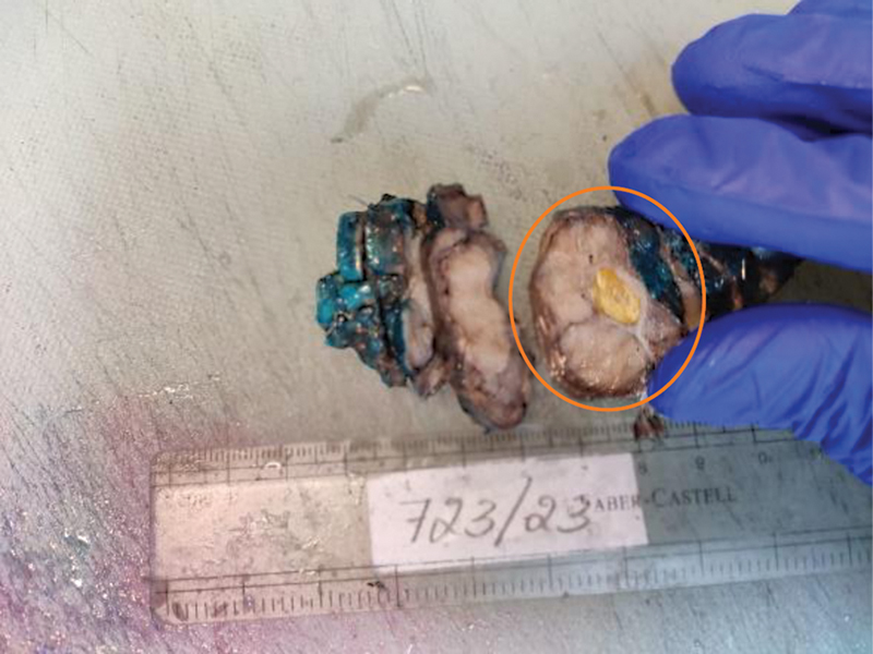
The patient underwent distal pancreatectomy. On gross examination and sectioning there was a well-circumscribed, fleshy oval nodule on the superior surface. Microscopic examination revealed pseudopapillae with hyalinized fibrovascular cores along with ossification and calcification. These findings were consistent with a SPT of the pancreas. Following resection the postoperative period was uneventful. The child was discharged and suggested regular follow-up ([Figs. 1] [2] [3] [4] [5] [6] [7] [8] [9] [10]).


Discussion
SPT of the pancreas is a rare exocrine tumor, described for the first time by Franz in 1959.[1] It is of low malignant potential and accounts for 1 to 2% of all exocrine pancreatic tumors,[2] but 52 to 71% of pancreatic tumors occur in children and adolescents.[3] [4]
Although SPT of the pancreas can affect patients at any age, most occur in young women in their second or third decade of life.[5] A large review reported that the mean age of patients with SPT of the pancreas is 21.97 years and that the male-to-female ratio is 1:9.78.[2]
If a pancreatic mass is detected in a child with no specific symptoms, it is likely to be a neuroendocrine tumor, solid and papillary epithelial neoplasm of the pancreas, or pancreatoblastoma.[6] A neuroendocrine tumor occurs at a slightly older age than solid and papillary epithelial neoplasm of the pancreas and has no female predilection. A neuroendocrine may appear cystic, contain calcification, demonstrate areas of internal hemorrhage, and metastasize to the liver.[7] [8] Pancreatoblastoma is a disease of childhood and has no female predilection. It is a more aggressive tumor than solid and papillary epithelial neoplasm of the pancreas and on diagnosis is frequently found to have liver metastasis. Radiologic studies have shown a sharply defined, round or lobulated heterogeneous mass, rarely with cystic spaces.[7] [8]
Clinical presentations of SPT of the pancreas range from asymptomatic “incidental” to unclear clinical features like abdominal pain or discomfort, poor appetite, and nausea, which are related to tumor compression on adjacent organs.[9]
SPT of the pancreas presenting with acute pancreatitis is not reported in literature. The case is a rare cause of acute pancreatitis.
The imaging features of SPT reflect the pathologic findings of cystic and solid components, intratumoral hemorrhage, a fibrous capsule, and, less commonly, calcifications.
On ultrasound, SPT presents as a well-circumscribed heterogeneous mass surrounded by a pseudocapsule of compressed pancreatic tissues and reactive fibrosis, sometimes with central cystic areas of necrosis.[10] The capsule may be visualized as an echogenic or, less commonly, hypoechoic rim at ultrasound and typically hypoattenuating at CT.[11] Unenhanced CT may identify hemorrhage and tiny calcifications.[10] [12]
MRI should be considered the best imaging technique for children due to the absence of radiation and its improved capacity for visualizing tumor components.[10] At MRI, T1-weighted images show a surrounding hypointense fibrous pseudocapsule and high signal intensity areas corresponding to internal hemorrhage, distinguishing features of SPT.[10] [12] Similar dark rim is also seen on T2-weighted images corresponding to the pseudocapsule.[2] The solid portions of the tumor are usually iso- to hypointense to pancreas on T1-weighted images and slightly heterogeneously hyperintense on T2-weighted images.[11] [12]
Recent studies indicate that these tumors may be classified into two subgroups—small (< 3 cm) and large (> 3 cm) SPTs, which have significantly different CT and MRI findings. Large SPTs (> 3 cm) are typically well demarcated, and encapsulated pancreatic lesions comprising both solid and cystic components. The cystic portions are usually central, whereas solid areas are commonly peripheral. These are typically hypoattenuating on unenhanced images and remain hypoattenuating during the pancreatic and portal venous phases. In contrast, the small (< 3 cm) SPTs are usually homogenous, predominantly solid tumors that may be ill-defined. On CT, these are hypoattenuating on unenhanced images, remain hypoattenuating on pancreatic phases, and may show homogeneous progressive enhancement to become isoattenuating on the hepatic venous phases.[13]
Aggressive features such as ductal dilatation, extracapsular invasion into adjacent structures including perineural and perivascular invasion, and nodal and distant metastases have been reported. The presence of a lobulated margin, focal discontinuity of the capsule, and pancreatic ductal dilation may suggest malignant SPT.
Surgical resection is the standard treatment for SPTs and is associated with excellent prognosis. Some authors have reported that male gender, elderly age group, tumor size > 5 cm, cellular or nuclear atypia, high mitotic rate, extensive necrosis, local invasion, and metastases are associated with poor prognosis.[14]
Conclusion
Solid pseudopapillary neoplasm of pancreas presenting in a male child with features of pancreatitis is rare. This case highlights how the imaging findings and clinical presentation of SPTs of the pancreas make it a difficult yet important diagnosis to reach. Knowledge that such tumors may mimic other pathologies can lead to a prompt workup and diagnosis.
Conflict of Interest
None declared.
-
References
- 1 Franz V. Papillary tumors of the pancreas: benign or malignant?. In: Frantz VK. ed. Atlas of Tumor Pathology. Washington, DC: U.S. Armed Forces Institute of Pathology; 1959: 32-33
- 2 Papavramidis T, Papavramidis S. Solid pseudopapillary tumors of the pancreas: review of 718 patients reported in English literature. J Am Coll Surg 2005; 200 (06) 965-972
- 3 Ahmed TS, Chavhan GB, Navarro OM, Traubici J. Imaging features of pancreatic tumors in children: 13-year experience at a pediatric tertiary hospital. Pediatr Radiol 2013; 43 (11) 1435-1443
- 4 Rojas Y, Warneke CL, Dhamne CA. et al. Primary malignant pancreatic neoplasms in children and adolescents: a 20 year experience. J Pediatr Surg 2012; 47 (12) 2199-2204
- 5 Soloni P, Cecchetto G, Dall'igna P, Carli M, Toffolutti T, Bisogno G. Management of unresectable solid papillary cystic tumor of the pancreas. A case report and literature review. J Pediatr Surg 2010; 45 (05) e1-e6
- 6 Cantisani V, Mortele KJ, Levy A. et al. MR imaging features of solid pseudopapillary tumor of the pancreas in adult and pediatric patients. AJR Am J Roentgenol 2003; 181 (02) 395-401
- 7 Jaksic T, Yaman M, Thorner P, Wesson DK, Filler RM, Shandling B. A 20-year review of pediatric pancreatic tumors. J Pediatr Surg 1992; 27 (10) 1315-1317
- 8 Buetow PC, Buck JL, Pantongrag-Brown L, Beck KG, Ros PR, Adair CF. Solid and papillary epithelial neoplasm of the pancreas: imaging-pathologic correlation on 56 cases. Radiology 1996; 199 (03) 707-711
- 9 Yu PF, Hu ZH, Wang XB. et al. Solid pseudopapillary tumor of the pancreas: a review of 553 cases in Chinese literature. World J Gastroenterol 2010; 16 (10) 1209-1214
- 10 Al-Qahtani S, Gudinchet F, Laswed T. et al. Solid pseudopapillary tumor of the pancreas in children: typical radiological findings and pathological correlation. Clin Imaging 2010; 34 (02) 152-156
- 11 Chung EM, Travis MD, Conran RM. Pancreatic tumors in children: radiologic-pathologic correlation. Radiographics 2006; 26 (04) 1211-1238
- 12 Moholkar S, Sebire NJ, Roebuck DJ. Solid-pseudopapillary neoplasm of the pancreas: radiological-pathological correlation. Pediatr Radiol 2005; 35 (08) 819-822
- 13 Ganeshan DM, Paulson E, Tamm EP, Taggart MW, Balachandran A, Bhosale P. Solid pseudo-papillary tumors of the pancreas: current update. Abdom Imaging 2013; 38 (06) 1373-1382
- 14 Lee JH, Yu J-S, Kim H. et al. Solid pseudopapillary carcinoma of the pancreas: differentiation from benign solid pseudopapillary tumour using CT and MRI. Clin Radiol 2008; 63 (09) 1006-1014
Address for correspondence
Publication History
Article published online:
15 May 2024
© 2024. The Author(s). This is an open access article published by Thieme under the terms of the Creative Commons Attribution License, permitting unrestricted use, distribution, and reproduction so long as the original work is properly cited. (https://creativecommons.org/licenses/by/4.0/)
Thieme Medical and Scientific Publishers Pvt. Ltd.
A-12, 2nd Floor, Sector 2, Noida-201301 UP, India
-
References
- 1 Franz V. Papillary tumors of the pancreas: benign or malignant?. In: Frantz VK. ed. Atlas of Tumor Pathology. Washington, DC: U.S. Armed Forces Institute of Pathology; 1959: 32-33
- 2 Papavramidis T, Papavramidis S. Solid pseudopapillary tumors of the pancreas: review of 718 patients reported in English literature. J Am Coll Surg 2005; 200 (06) 965-972
- 3 Ahmed TS, Chavhan GB, Navarro OM, Traubici J. Imaging features of pancreatic tumors in children: 13-year experience at a pediatric tertiary hospital. Pediatr Radiol 2013; 43 (11) 1435-1443
- 4 Rojas Y, Warneke CL, Dhamne CA. et al. Primary malignant pancreatic neoplasms in children and adolescents: a 20 year experience. J Pediatr Surg 2012; 47 (12) 2199-2204
- 5 Soloni P, Cecchetto G, Dall'igna P, Carli M, Toffolutti T, Bisogno G. Management of unresectable solid papillary cystic tumor of the pancreas. A case report and literature review. J Pediatr Surg 2010; 45 (05) e1-e6
- 6 Cantisani V, Mortele KJ, Levy A. et al. MR imaging features of solid pseudopapillary tumor of the pancreas in adult and pediatric patients. AJR Am J Roentgenol 2003; 181 (02) 395-401
- 7 Jaksic T, Yaman M, Thorner P, Wesson DK, Filler RM, Shandling B. A 20-year review of pediatric pancreatic tumors. J Pediatr Surg 1992; 27 (10) 1315-1317
- 8 Buetow PC, Buck JL, Pantongrag-Brown L, Beck KG, Ros PR, Adair CF. Solid and papillary epithelial neoplasm of the pancreas: imaging-pathologic correlation on 56 cases. Radiology 1996; 199 (03) 707-711
- 9 Yu PF, Hu ZH, Wang XB. et al. Solid pseudopapillary tumor of the pancreas: a review of 553 cases in Chinese literature. World J Gastroenterol 2010; 16 (10) 1209-1214
- 10 Al-Qahtani S, Gudinchet F, Laswed T. et al. Solid pseudopapillary tumor of the pancreas in children: typical radiological findings and pathological correlation. Clin Imaging 2010; 34 (02) 152-156
- 11 Chung EM, Travis MD, Conran RM. Pancreatic tumors in children: radiologic-pathologic correlation. Radiographics 2006; 26 (04) 1211-1238
- 12 Moholkar S, Sebire NJ, Roebuck DJ. Solid-pseudopapillary neoplasm of the pancreas: radiological-pathological correlation. Pediatr Radiol 2005; 35 (08) 819-822
- 13 Ganeshan DM, Paulson E, Tamm EP, Taggart MW, Balachandran A, Bhosale P. Solid pseudo-papillary tumors of the pancreas: current update. Abdom Imaging 2013; 38 (06) 1373-1382
- 14 Lee JH, Yu J-S, Kim H. et al. Solid pseudopapillary carcinoma of the pancreas: differentiation from benign solid pseudopapillary tumour using CT and MRI. Clin Radiol 2008; 63 (09) 1006-1014