

Subscribe to RSS
DOI: 10.1055/s-0044-1791234
The Effects of Constant Light and Running-Wheel Access in Middle-Aged Female C57BL6/J Mice
Authors
Funding Source The author(s) received no financial support for the research.

Abstract
Objective Chronic light exposure is associated with poorer mental and physical health. Many groups are chronically exposed to light due to their work schedule, including night-shift nurses. The present study aims to determine if middle-aged female mice have an increased vulnerability to chronic light exposure compared with younger individuals.
Materials and Methods Young and middle-age female mice were housed in cages with or without running wheels and were exposed to either a 12:12-hour light:dark cycle or constant light (LL). All mice were assayed for circadian locomotor activity and anxiety-like behaviors, and weight, food consumption, and estrogen receptor beta (ERβ) levels were measured.
Results Middle-aged mice exhibited longer circadian periods in LL, reduced circadian power, and increased food consumption compared with young mice. LL increased novelty-induced activity and reduced ERβ regardless of age.
Discussion These results indicate that middle-aged females may have an increased susceptibility to the negative circadian consequences caused by constant bright light exposure.
Introduction
Chronic exposure to light during the night can have several negative behavioral and physiological effects on individuals. Exposure to light at night can lead to irregular sleep patterns, impaired cognitive function (attention, memory, and decision-making), and mood disturbances (such as irritability, anxiety, and depression).[1] Additionally, aberrant light exposure has been linked[2] to an increased risk of metabolic disorders, such as obesity and diabetes. Exposure to light during nighttime hours may also alter the behavioral cycles of individuals; this leads to changes in eating patterns and social interactions, which, affect the regulation of emotionality and metabolism, resulting in a misalignment with natural timing cues.
Certain factors associated with aging can influence the timing and synchronization of circadian rhythms. In older humans, there is a tendency for circadian rhythms to phase advance, exhibiting a shift toward an earlier chronotype as well as a shortening of their free-running rhythm.[3] Interestingly, studies investigating how aging affects the circadian period in animal models are somewhat variable depending on the rodent used. In constant darkness (DD), rats and hamsters tend to experience period shortening as they age,[4] [5] [6] while mice exhibit period lengthening.[7] [8] [9] Despite numerous studies on the effects of aging in standard light:dark (LD) cycles and DD, few studies have investigated how animals and humans of differing ages respond to chronic light exposure.
In young animal models, estrogen provides a protective effect from circadian disruption, preventing negative metabolic and rhythmic outcomes.[10] [11] Estrogen receptors (ERs) are also known to regulate circadian rhythmicity via direct modulation of core clock genes within the suprachiasmatic nucleus (SCN; as reviewed by Hatcher [et al.12]), and a recent study[13] showed that light at night can produce reductions in ER beta (ERβ). However, aging may produce reductions in both estrogen and ER levels (as reviewed by Maioli [et al.14]), which may explain the negative effects of aging on the robustness of circadian rhythms. As such, middle-aged and older women may be more susceptible to circadian disruption compared with younger women, and, in some cases, may be more likely to be exposed to light at night due to work or other societal demands. One study[15] reported that, out of almost 55 thousand female nurses surveyed in the United States, 17% of those aged 36–64 years worked only night shifts. Regarding mouse studies, many of them tend to skew their investigations toward younger mice (aged between 6 and 20 weeks), despite the fact that many physiological changes occur in this stage of their lives.[16] As most studies use mice younger than 6 months of age and given that many middle-aged nurses work the night shift, research that investigates these parameters would be important for these individuals and for their health outcomes. The present study investigated whether middle-aged female mice (∼ 50 weeks old by the end of the experiment) present different behavioral and physiological responses to constant light (LL) exposure compared with younger mice (∼ 22 weeks old by the end of the experiment).
Materials and Methods
All experiments listed below were approved by the Bridgewater State University's Institutional Animal Care and Use Committee.
Animals and Circadian Monitoring
In total, 67 C57BL/6J female mice (B6J; strain number 000664; The Jackson Laboratory, Bar Harbor, ME, United States) were purchased; 34 of them were young (Y), aged 8 weeks, and 33 were middle-aged (MA), with ages ranging from 35 to 38 weeks. Approximately half of the Y group and half of the MA group were housed individually in circadian activity cages monitored through infrared (IR) beams, and the other halves, in running wheel (RW) cages (Starr Life Sciences Corp., Oakmont, PA, United States), as previously described by Nascimento et al.,[17] at 22°C under 150-lux lighting (long light-emitting diodes [LEDs] embedded in the ceiling). All animals were initially placed in a 12:12-hour LD cycle with water and rodent chow (Laboratory Rodent Diet 5001, Land O'Lakes, Inc., Arden Hills, MN, United States). After 1 week of entrainment and acclimation, half of the Y group in IR and RW cages and half of the MA group in IR and RW cages were exposed to LL (150 lux), while the remaining mice stayed in the standard 12:12-hour LD cycle. As such, there were 8 groups: 1) LD/MA/IR (n = 8); 2) LD/Y/IR (n = 8); 3) LL/MA/IR (n = 8); 4) LL/Y/IR (n = 8); 5) LD/MA/RW (n = 8); 6) LD/Y/RW (n = 9); 7) LL/MA/RW (n = 9); and 8) LL/Y/RW (n = 9). Weight and food consumption were measured weekly. All of the assays listed below were conducted during the middle of each mouse's inactive time (approximately Zeitgeber time [ZT] 6 for LD animals and circadian time [CT] 6 for LL animals) and in the light to form the basis of comparison.
Behavioral Assays
All mice were subjected to open-field and light-dark box assays after 10 and 11 weeks of exposure to their respective light cycle, as previously described by Hicks et al.[18]
ERβ Enzyme-linked Immunosorbent Assays
One week after the completion of the light-dark box assay, each mouse was euthanized via CO2 narcosis, and their whole brains were immediately stored at -80°C. Their SCNs (identified by their location at the base of the third ventricle, with the medial optic chiasm as a point of reference) were then removed using a dissecting microscope, as previously described by Michaud et al.[13] Homogenates were created in a cocktail containing Pierce IP lysis buffer (Thermo Fisher Scientific, Waltham, MA, United States) and Halt protease inhibitor cocktail, EDTA-Free (100 × ) (Thermo Fisher Scientific) at a ratio of 100 μL of protease inhibitor for each 10 mL of lysis buffer, and a ratio of 0.2 mL of protease/lysis cocktail was added to 5 mg of SCN sample. The supernatant was used in mouse ERβ enzyme-linked immunosorbent assays (ELISAs; MBS776119, MyBioSource, Inc., San Diego, CA, United States).
Statistical Analyses
Circadian period (estimated through the Chi-square periodogram) and daily locomotor activity were calculated using the ClockLab software (Actimetrics, Lafayette, IN, United States). Three-way analysis of variance (ANOVA) with Tukey post-hoc pairwise comparisons was (age versus cage versus light cycle) used to uncover mean differences among the groups regarding all dependent variables.
Results
Circadian Locomotor Activity
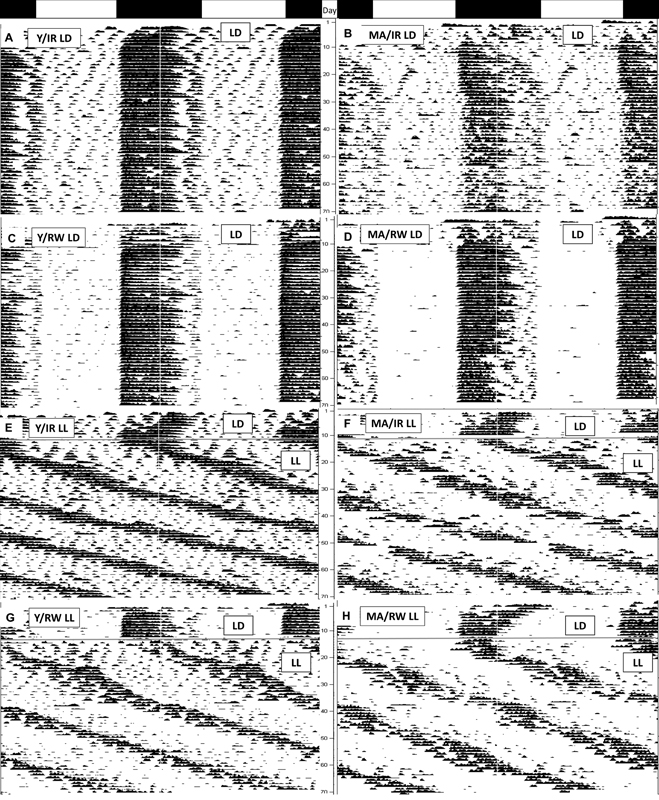
Representative actograms are shown in [Fig. 1]. All animals were able to entrain to the 12:12 LD cycle, and all animals in LL remained rhythmic throughout the entirety of the experiment. One animal (LD/Y/RW) was excluded due to a malfunction in the wire of the activity monitor. As expected, mice in LL experienced period lengthening (F1,59 = 31.68; p < 0.001). A three-way interaction was found for the circadian period (F1,59 = 4.44; p = 0.040). In LL, MA/IR mice had longer free-running periods compared with Y/IR mice (p = 0.029), while MA/RW mice exhibited similar circadian periods compared with Y/RW mice (p = 0.99). Additionally in LL, while Y/IR and Y/RW mice exhibited similar period lengths in LL (p = 0.33), MA/IR mice showed increased period lengthening compared with MA/RW mice (p < 0.001) ([Fig. 2A]). Age (F1,59 = 10.15; p = 0.002; Y > MA) and light cycle (F1,59 = 40.70; p < 0.001; LD > LL) affected circadian power, but cage type produced no changes (p = 0.14) ([Fig. 2B]). Light cycle (F1,59 = 21.07; p < 0.001) and cage type (F1,59 = 9.59; p = 0.003) affected daily locomotor activity. A three-way interaction was also uncovered for daily activity levels (F1,59 = 4.48; p = 0.039): whereas LD/RW/Y mice exhibited increase when running compared with LL/RW/Y mice (p = 0.006), RW/MA mice had no such difference between light cycles (p = 0.98) ([Fig. 2C]).

Anxiety-like Behavioral Assays
The responses to both open-field and light-dark box assays are presented in [Table 1]. For the open-field assay, LL increased distance traveled (F1,60 = 9.10; p = 0.005), velocity (F1,60 = 4.97; p = 0.030), and total rotations (F1,60 = 5.17; p = 0.027). Additionally, RW animals also exhibited a reduction in the distance traveled (F1,60 = 6.27; p = 0.015), but the age of the animal had no effect on this behavior (p = 0.44). A cage/age interaction was observed for velocity (F1,60 = 4.77; p = 0.033): whereas MA/IR mice exhibited increased velocity compared with MA/RW mice (p = 0.023), Y/IR and Y/RW mice exhibited no difference in velocity (p = 0.99). No differences were observed for rears (p = 0.072) and center zone time (p = 0.067). For the light-dark box, one mouse (LD/Y/IR) was excluded, as it did not make one transition into the dark zone. No differences were observed among the groups regarding time spent in the dark zone (p = 0.31). Light cycle (F1,59 = 4.28; p = 0.044; LD < LL) and cage type (F1,59 = 18.35; p < 0.001; IR > RW) influenced the number of transitions between the light and dark zones, although the age of the animal produced no changes in transitions between the two zones (p = 0.54).
Abbreviations: LD, light:dark; LL, constant light; IR, infrared; MA, middle-aged; RW, running wheel; Y, young.
Notes: Constant light increased acute locomotor activity within both the open-field and light-dark box assays, as measured by distance traveled, velocity, and number of rotations in the open-field assay and increased transitions in the light-dark box assay. Animals with a running wheel also exhibited reduced locomotor activity in these assays, as measured by distance traveled and velocity in the open-field assay and decreased transitions in the light-dark box assay. Values with different letters (a, b, c, d) indicate significantly from each other at p ≤ 0.050.
Physiological Measurements
A cycle/age interaction was found regarding weight gain (F1,60 = 5.97; p = 0.018): while Y mice gained more weight compared with MA mice in LD (p = 0.001), no differences were found between mice of these two age groups in LL (p = 0.80) ([Fig. 3A]). Cage type had no effect on weight gain (p = 0.58). Overall, light cycle (F1,60 = 5.82; p = 0.019; LD > LL), RW access (F1,60 = 30.81; p < 0.001; RW > IR) and age (F1,60 = 24.69; p = 0.001; MA > Y) altered food intake. A cage/age interaction was observed regarding the average weekly food consumption (F1,60 = 7.35; p = 0.009): While IR mice consumed similar amounts of food regardless of age (p = 0.41), MA/RW mice exhibited increased food consumption compared with Y/RW mice (p < 0.001). Additionally, while no differences were found between Y/IR and Y/RW mice (p = 0.19), MA/IR mice consumed less food per week than MA/RW mice (p < 0.001) ([Fig. 3B]). Exposure to LL reduced ERβ within the SCN (F1,52 = 17.19; p < 0.001) ([Fig. 3C]). Age (p = 0.20) and cage type (p = 0.22) had no effects on ERβ.

Discussion
The present study reports two differences in circadian behavior between Y and MA female B6 mice when exposed to LL. First, MA/IR mice exhibited lengthened free running rhythms in LL compared with Y mice. This result corroborates those of previous studies[7] [8] [9] [19] which report that aging in mice can lead to increased period lengths in DD, although this effect seems to taper off once “middle-age” has passed (∼ 15–18 months of age). While previous research[3] [4] [5] [6] [7] shows that aging can affect the free running period of the circadian clock, their result on how the period changes can vary depending on species and genetic model used. Conversely, humans, rats, and hamsters tend to shorten their period lengths as they age in DD,[3] [4] [5] [6] [20] and this shortening coincides with shortened SCN rhythms in period1:luciferase rats.[21] This difference may be due to the delay/advance ratios inherent in each species due to their endogenous period lengths. Mice tend to have rhythms shorter than 24 hours while humans, rats, and hamsters tend to have rhythms longer than 24 hours, so aging would seem to disrupt the functioning of the clock by having the rhythm trend in the “other direction,” but this possibility has not yet been directly tested. It is also worth noting that some aging studies (including studies conducted by different authors in the same laboratory) report no differences in free running period lengths in aged animals, including humans.[22] [23] [24] Even though some studies observe circadian period changes and others do not, in those that do, the direction of the period change during the aging process (mice lengthening versus rats/hamsters/humans shortening) is consistent.
While numerous studies[6] [7] have investigated the effects of aging in DD conditions, few studies have evaluated the circadian responses of old individuals to chronic light exposure. Interestingly, two of the studies[25] [26] that investigated the effects of aging in LL used the period2:luciferase construct. In DD, the free running period of both locomotor activity and SCN explants of PER2-LUC mice lengthen as they aged.[8] However, the rhythmic outcomes of aging on LL in PER2-LUC mice are somewhat unusual, for individual mice can exhibit either a period lengthening or a period shortening to under 24 hours.[25] [26] This atypical result of period shortening in LL may be due to alterations in circadian rhythmicity and molecular mechanisms inherent in the genetic construct. Compared with wild-type mice, PER2-LUC mice exhibit altered phase shifting behavior and PER2 turnover,[26] as well as increased photoreceptor degeneration as they age.[27] Aged wild-type Syrian hamsters exhibit reduced period lengthening in LL compared with young hamsters.[4] The period lengthening observed in this study using wild-type mice and in hamsters in LL[4] mirrors what is usually observed in DD for aged rodents. Additional studies using mice, rats, and hamsters will be extremely useful in determining how species with different endogenous periods respond to LL as they age.
In the present study, MA mice exhibited reduced circadian power compared with Y mice in both LD and LL. This result seems to be directly related to aging, as numerous studies[5] [8] also demonstrate reduced circadian robustness in animal models as they age. For example, fetal SCN transplants to old mice are sufficient to restore the robustness of the circadian rhythm in both LD and DD in rats.[28] Regardless of their direction in period change, mPER2-LUC rhythms in LD, LL, and DD dampen quicker in aged versus Y PER2-LUC mice.[8] [25] Part of these disruptions to circadian power may be due to reductions in light-sensitive proteins that control input signals to the SCN. Vasoactive intestinal peptide (VIP), a light-harvesting protein within the SCN, is markedly reduced in both MA and old rodents[29] [30] (although the effect is not as large in MA animals compared with older ones). Additionally, both LL[31] and suppression of VIP can lead to aging-like consequences.[32] Elderly mice also have decreased circadian photosensitivity compared with younger mice due to a reduction in N-methyl-D-aspartate (NMDA) receptors that contain subunit 2B.[33] As such, we initially hypothesized that the middle-aged mice in our study may have exhibited reduced period lengthening in LL due to their reduction in photosensitivity and circadian power, as seen in other models of reduced photosensitivity and reduced light-harvesting proteins,[34] [35] but the results from this study to not fit with that supposition. Rather, aging might lead to a quickening of SCN neurons decoupling from each other,[8] an outcome sometimes observed under LL.[36] In addition to reductions in VIP, aging also leads to declines in gamma-aminobutyric acid-ergic (GABAergic) signaling within the SCN – GABA is a neurotransmitter that synchronizes SCN neurons.[37] Additionally, a recent study[38] mathematically modeling aging in mice determined that weaker oscillations due to decoupling of SCN networks is associated with longer periods – and period lengthening is an outcome usually observed in aging mice. The results of the present study also show that MA/RW mice do not exhibit period lengthening to the extent observed among MA/IR mice. Interestingly, exercise can prevent the phase decoupling effects of aging and LL in both the SCN[39] and peripheral tissues[40] without improving circadian power, even in PER2-LUC mice. These results indicate that chronic exercise may act as a countermeasure to prevent the circadian decoupling issues associated with aging. As such, the change in the timing of the rhythm sometimes observed with aging may be a consequence of SCN decoupling and weaker oscillatory networks, which may alter the free running period in the direction away from each species endogenous period length (that is, aging lengthens rhythms in shorter than 24-hour mice and shortens rhythms in longer than 24-hour rats/hamsters/humans). The mechanism that connects these age-related changes in circadian robustness and period have not yet been fully elucidated. All the same, while the effects of aging on the circadian period seem to vary based on species and experimental protocol, the reduction in circadian robustness during the aging process is consistent across species and studies.
Another possible explanation as to why age alters the power and period of the clock may be due to alterations in gonadal hormone receptors within the brain. Reductions in gonadal hormones and receptors influence both the period and robustness of the circadian clock,[10] [13] and aging reduces the levels of ERs, including ERβ, within the hypothalamus.[14] Estrogen receptor beta is downregulated by period,[41] and as LL increases period expression within the SCN, this may be why nightly exposure to light alters ERs. It is worth noting that neither period transcripts nor estrogen binding were directly measured in the current study. As the reduction in ERβ caused by LL was similar among the groups, the differences in circadian periodicity and power between MA and Y mice in LL for the present study may not be due to alterations in ERs.
Despite the differences in chronic circadian locomotor activity, acute/exploratory locomotor activity and anxiety-like behaviors in the open-field and light-dark box assays were largely unaffected by age. We were able to replicate the recent findings that LL increases novelty-induced locomotor activity regardless of sex (that is, increased activity in the open-field assay and transitions in the light-dark box assay) in mouse strains with high baseline levels of locomotor activity, including B6J.[10] [42] This novelty-induced hyperactivity could be an expression of increased darting or rapid movement within the novel environment, and interpreted as increased stress, impulsivity or manic behavior, as observed in both human and animal models when exposed to chronic light.[43] [44] Estrogen receptor beta is known to regulate mood as mouse studies report ER knockout mice exhibit increases to those behaviors in behavioral assays compared with controls,[45] [46] so some of the changes in locomotor activity observed in the open-field and light-dark box assays in the present study may be attributed to the reduction in ERβ. The current study also replicates a well-known finding that RW access reduces acute novelty-induced locomotor activity in the open-field and light-dark box assays (see “Discussion” sections in the studies by Hicks et al.[18] and Capri et al.[42]), even in mice up to 15 months old.[47] Wheel-running is considered a rewarding activity, modeling a form of voluntary exercise, which in rodent studies usually produces antidepressive and antianxiety effects.[48] The results seen here mirror what is found in pharmacological studies using anxiolytics in mice.[49] [50] Whether nightly exposure to light or wheel-running affects mice older than 15 months (that is, past middle-age) needs to be further explored.
On the other hand, Y mice experience increased weight gain compared with MA mice in LD only. Overall, MA mice in LD gained little weight on average during the experiment, while in LL they gained a moderate amount, similar to that of Y mice. As the Y mice gained similar weight regardless of their cage type in both LD and LL, this result indicates that the effects of LL on body weight regulation can be altered as animals age. In fact, the MA/LD mice gained little weight, well within the possible margin of daily fluctuations, since a previous study[51] reported that body mass in female mice can fluctuate almost as much as 1.5 g daily due to variations in food consumption throughout the day. For the Y mice, this weight gain is not only due to variations in the phase of the collection time, it is also the weight gain expected in young animals as they mature toward older adulthood. This result is in line with reports that Y (6 months or younger) male and female (even ovariectomized) B6J mice tend not to gain weight in LL compared with other B6 substrains, such as B6N.[10] [42] It is worth noting that food consumption was reduced overall in LL and higher among MA mice. As Y/LD mice presented the most weight gain, this result suggests that food intake had little relationship to weight gain in the present study. The difference between RW and IR mice in terms of weight gain is not due to differences in food intake among MA mice, as MA/RW mice in LD exhibited increased food intake and did not gain much weight.
One caveat to consider is that the results presented in the current study reflect the analysis of behavioral and physiological outcomes from a single time point (middle of the day) during the circadian cycle. While the circadian locomotor behavior of the animals was in phase, other physiological rhythms may be out of alignment between the LD and LL groups and the Y and MA groups, which may be the case considering how weight gain was not correlated with food intake in the present study. Nevertheless, the current study was able to replicate the behavioral effects of LL and wheel-running during the subjective day, which led to increased and reduced novelty-induced activity respectively in rodent models.
In conclusion, we have shown that MA and Y mice experience different behavioral and physiological responses to LL and RW access. Middle-aged mice exhibit different responses in terms of their circadian rhythmicity and robustness compared with Y mice. The present study adds to the body of evidence that mice tend to lengthen their circadian period as they age, and that chronic light exposure, regardless of aging, has negative effects on behavioral and physiological outcomes. Studying aging in the context of nightshift work and nightly exposure to light is important, as it involves the intersection of circadian rhythms, sleep patterns, behavioral outcomes, safety, and overall health. It is essential to develop strategies to mitigate the potential negative consequences of night work and promote good light and sleep hygiene among individuals engaged in such work schedules or among individuals exposed to high amounts of light pollution.
Conflict of Interests
The authors have no conflict of interests to declare.
-
References
- 1 Tancredi S, Urbano T, Vinceti M, Filippini T. Artificial light at night and risk of mental disorders: A systematic review. Sci Total Environ 2022; 833: 155185
- 2 Fonken LK, Workman JL, Walton JC. et al. Light at night increases body mass by shifting the time of food intake. Proc Natl Acad Sci U S A 2010; 107 (43) 18664-18669
- 3 Weitzman ED, Moline ML, Czeisler CA, Zimmerman JC. Chronobiology of aging: temperature, sleep-wake rhythms and entrainment. Neurobiol Aging 1982; 3 (04) 299-309
- 4 Morin LP. Age, but not pineal status, modulates circadian periodicity of golden hamsters. J Biol Rhythms 1993; 8 (03) 189-197
- 5 Penev PD, Zee PC, Turek FW. Quantitative analysis of the age-related fragmentation of hamster 24-h activity rhythms. Am J Physiol 1997; 273 (06) R2132-R2137
- 6 Witting W, Mirmiran M, Bos NP, Swaab DF. The effect of old age on the free-running period of circadian rhythms in rat. Chronobiol Int 1994; 11 (02) 103-112
- 7 Possidente B, McEldowney S, Pabon A. Aging lengthens circadian period for wheel-running activity in C57BL mice. Physiol Behav 1995; 57 (03) 575-579
- 8 Nakamura TJ, Nakamura W, Tokuda IT. et al. Age-Related Changes in the Circadian System Unmasked by Constant Conditions. eNeuro 2015;2(04):ENEURO.0064-15.2015
- 9 Valentinuzzi VS, Scarbrough K, Takahashi JS, Turek FW. Effects of aging on the circadian rhythm of wheel-running activity in C57BL/6 mice. Am J Physiol 1997; 273 (06) R1957-R1964
- 10 Michaud JM, Price JC, Deane HV. et al. The effects of ovariectomy on the behavioral and physiological responses to constant light in C57BL6/J Mice. Biol Rhythm Res 2022; 53 (06) 921-938
- 11 Zhu L, Zou F, Yang Y. et al. Estrogens prevent metabolic dysfunctions induced by circadian disruptions in female mice. Endocrinology 2015; 156 (06) 2114-2123
- 12 Hatcher KM, Royston SE, Mahoney MM. Modulation of circadian rhythms through estrogen receptor signaling. Eur J Neurosci 2020; 51 (01) 217-228
- 13 Michaud JM, Waring CT, Medeiros Contini F. et al. Estradiol Regulates Circadian Responses to Acute and Constant Light Exposure in Female Mice. J Biol Rhythms 2023; 38 (04) 407-415
- 14 Maioli S, Leander K, Nilsson P, Nalvarte I. Estrogen receptors and the aging brain. Essays Biochem 2021; 65 (06) 913-925
- 15 Ramin C, Devore EE, Wang W, Pierre-Paul J, Wegrzyn LR, Schernhammer ES. Night shift work at specific age ranges and chronic disease risk factors. Occup Environ Med 2015; 72 (02) 100-107
- 16 Jackson SJ, Andrews N, Ball D. et al. Does age matter? The impact of rodent age on study outcomes. Lab Anim 2017; 51 (02) 160-169
- 17 Nascimento NF, Hicks JA, Carlson KN, Hatzidis A, Amaral DN, Seggio JA. 6-h advances alter circadian activity patterns, fasting glucose, and insulin levels in C57BL6/J mice. Biol Rhythm Res 2016; 47 (01) 133-143
- 18 Hicks JA, Hatzidis A, Arruda NL. et al. Voluntary wheel-running attenuates insulin and weight gain and affects anxiety-like behaviors in C57BL6/J mice exposed to a high-fat diet. Behav Brain Res 2016; 310: 1-10
- 19 Banks G, Heise I, Starbuck B. et al. Genetic background influences age-related decline in visual and nonvisual retinal responses, circadian rhythms, and sleep. Neurobiol Aging 2015; 36 (01) 380-393
- 20 Kendall AR, Lewy AJ, Sack RL. Effects of aging on the intrinsic circadian period of totally blind humans. J Biol Rhythms 2001; 16 (01) 87-95
- 21 Yamazaki S, Straume M, Tei H, Sakaki Y, Menaker M, Block GD. Effects of aging on central and peripheral mammalian clocks. Proc Natl Acad Sci U S A 2002; 99 (16) 10801-10806
- 22 Kolker DE, Vitaterna MH, Fruechte EM, Takahashi JS, Turek FW. Effects of age on circadian rhythms are similar in wild-type and heterozygous Clock mutant mice. Neurobiol Aging 2004; 25 (04) 517-523
- 23 Duffy JF, Viswanathan N, Davis FC. Free-running circadian period does not shorten with age in female Syrian hamsters. Neurosci Lett 1999; 271 (02) 77-80
- 24 Czeisler CA, Duffy JF, Shanahan TL. et al. Stability, precision, and near-24-hour period of the human circadian pacemaker. Science 1999; 284 (5423) 2177-2181
- 25 Polidarová L, Sládek M, Novosadová Z, Sumová A. Aging does not compromise in vitro oscillation of the suprachiasmatic nuclei but makes it more vulnerable to constant light. Chronobiol Int 2017; 34 (01) 105-117
- 26 Ralph MR, Shi SQ, Johnson CH. et al. Targeted modification of the Per2 clock gene alters circadian function in mPer2luciferase (mPer2Luc) mice. PLOS Comput Biol 2021; 17 (05) e1008987
- 27 Goyal V, DeVera C, Baba K. et al. Photoreceptor Degeneration in Homozygous Male Per2luc Mice During Aging. J Biol Rhythms 2021; 36 (02) 137-145
- 28 Li H, Satinoff E. Fetal tissue containing the suprachiasmatic nucleus restores multiple circadian rhythms in old rats. Am J Physiol 1998; 275 (06) R1735-R1744
- 29 Duncan MJ, Herron JM, Hill SA. Aging selectively suppresses vasoactive intestinal peptide messenger RNA expression in the suprachiasmatic nucleus of the Syrian hamster. Brain Res Mol Brain Res 2001; 87 (02) 196-203
- 30 Krajnak K, Kashon ML, Rosewell KL, Wise PM. Aging alters the rhythmic expression of vasoactive intestinal polypeptide mRNA but not arginine vasopressin mRNA in the suprachiasmatic nuclei of female rats. J Neurosci 1998; 18 (12) 4767-4774
- 31 Vinogradova IA, Anisimov VN, Bukalev AV. et al. Circadian disruption induced by light-at-night accelerates aging and promotes tumorigenesis in young but not in old rats. Aging (Albany NY) 2010; 2 (02) 82-92
- 32 Gerhold LM, Rosewell KL, Wise PM. Suppression of vasoactive intestinal polypeptide in the suprachiasmatic nucleus leads to aging-like alterations in cAMP rhythms and activation of gonadotropin-releasing hormone neurons. J Neurosci 2005; 25 (01) 62-67
- 33 Biello SM, Bonsall DR, Atkinson LA, Molyneux PC, Harrington ME, Lall GS. Alterations in glutamatergic signaling contribute to the decline of circadian photoentrainment in aged mice. Neurobiol Aging 2018; 66: 75-84
- 34 Deane HV, Concepcion HA, Gatewood AE, Quintana J, Seggio JA. Strain specific behavioral and physiological responses to constant light in male CBA/J and CBA/CaJ mice. Sleep Sci 2021; 14 (Spec 2): 167-173
- 35 Ruby NF, Brennan TJ, Xie X. et al. Role of melanopsin in circadian responses to light. Science 2002; 298 (5601) 2211-2213
- 36 Ohta H, Yamazaki S, McMahon DG. Constant light desynchronizes mammalian clock neurons. Nat Neurosci 2005; 8 (03) 267-269
- 37 Palomba M, Nygård M, Florenzano F, Bertini G, Kristensson K, Bentivoglio M. Decline of the presynaptic network, including GABAergic terminals, in the aging suprachiasmatic nucleus of the mouse. J Biol Rhythms 2008; 23 (03) 220-231
- 38 Myung J, Hong S, Schmal C, Vitet H, Wu MY. Weak synchronization can alter circadian period length: implications for aging and disease conditions. Front Neurosci 2023; 17: 1242800
- 39 Leise TL, Harrington ME, Molyneux PC. et al. Voluntary exercise can strengthen the circadian system in aged mice. Age (Dordr) 2013; 35 (06) 2137-2152
- 40 Hamaguchi Y, Tahara Y, Hitosugi M, Shibata S. Impairment of Circadian Rhythms in Peripheral Clocks by Constant Light Is Partially Reversed by Scheduled Feeding or Exercise. J Biol Rhythms 2015; 30 (06) 533-542
- 41 Cai W, Rambaud J, Teboul M. et al. Expression levels of estrogen receptor beta are modulated by components of the molecular clock. Mol Cell Biol 2008; 28 (02) 784-793
- 42 Capri KM, Maroni MJ, Deane HV. et al. Male C57BL6/N and C57BL6/J Mice Respond Differently to Constant Light and Running-Wheel Access. Front Behav Neurosci 2019; 13: 268
- 43 Byun JI, Lee BU, Koo YS. et al. Bright light exposure before bedtime impairs response inhibition the following morning: a non-randomized crossover study. Chronobiol Int 2018; 35 (08) 1035-1044
- 44 Li Z, Lee CS, Chen S. et al. Blue light at night produces stress-evoked heightened aggression by enhancing brain-derived neurotrophic factor in the basolateral amygdala. Neurobiol Stress 2023; 28: 100600
- 45 Lund TD, Rovis T, Chung WC, Handa RJ. Novel actions of estrogen receptor-beta on anxiety-related behaviors. Endocrinology 2005; 146 (02) 797-807
- 46 Imwalle DB, Gustafsson JA, Rissman EF. Lack of functional estrogen receptor beta influences anxiety behavior and serotonin content in female mice. Physiol Behav 2005; 84 (01) 157-163
- 47 Morgan JA, Singhal G, Corrigan F, Jaehne EJ, Jawahar MC, Baune BT. The effects of aerobic exercise on depression-like, anxiety-like, and cognition-like behaviours over the healthy adult lifespan of C57BL/6 mice. Behav Brain Res 2018; 337: 193-203
- 48 Duman CH, Schlesinger L, Russell DS, Duman RS. Voluntary exercise produces antidepressant and anxiolytic behavioral effects in mice. Brain Res 2008; 1199: 148-158
- 49 Clément Y, Prut L, Saurini F. et al. Gabra5-gene haplotype block associated with behavioral properties of the full agonist benzodiazepine chlordiazepoxide. Behav Brain Res 2012; 233 (02) 474-482
- 50 Quines CB, Da Rocha JT, Sampaio TB. et al. Involvement of the serotonergic system in the anxiolytic-like effect of 2-phenylethynyl butyltellurium in mice. Behav Brain Res 2015; 277: 221-227
- 51 Kawamura S, Yamazoe H, Hosokawa Y. Diurnal Gain and Nocturnal Reduction of Body Weight in Young Adult Rabbits: The Reverse of the Circadian Rhythm Observed in Rats and Mice. J Toxicol Cur Res 2020; 4: 16
Address for correspondence
Publication History
Received: 15 January 2024
Accepted: 13 August 2024
Article published online:
08 May 2025
© 2025. Brazilian Sleep Association. This is an open access article published by Thieme under the terms of the Creative Commons Attribution-NonDerivative-NonCommercial License, permitting copying and reproduction so long as the original work is given appropriate credit. Contents may not be used for commercial purposes, or adapted, remixed, transformed or built upon. (https://creativecommons.org/licenses/by-nc-nd/4.0/)
Thieme Revinter Publicações Ltda.
Rua do Matoso 170, Rio de Janeiro, RJ, CEP 20270-135, Brazil
-
References
- 1 Tancredi S, Urbano T, Vinceti M, Filippini T. Artificial light at night and risk of mental disorders: A systematic review. Sci Total Environ 2022; 833: 155185
- 2 Fonken LK, Workman JL, Walton JC. et al. Light at night increases body mass by shifting the time of food intake. Proc Natl Acad Sci U S A 2010; 107 (43) 18664-18669
- 3 Weitzman ED, Moline ML, Czeisler CA, Zimmerman JC. Chronobiology of aging: temperature, sleep-wake rhythms and entrainment. Neurobiol Aging 1982; 3 (04) 299-309
- 4 Morin LP. Age, but not pineal status, modulates circadian periodicity of golden hamsters. J Biol Rhythms 1993; 8 (03) 189-197
- 5 Penev PD, Zee PC, Turek FW. Quantitative analysis of the age-related fragmentation of hamster 24-h activity rhythms. Am J Physiol 1997; 273 (06) R2132-R2137
- 6 Witting W, Mirmiran M, Bos NP, Swaab DF. The effect of old age on the free-running period of circadian rhythms in rat. Chronobiol Int 1994; 11 (02) 103-112
- 7 Possidente B, McEldowney S, Pabon A. Aging lengthens circadian period for wheel-running activity in C57BL mice. Physiol Behav 1995; 57 (03) 575-579
- 8 Nakamura TJ, Nakamura W, Tokuda IT. et al. Age-Related Changes in the Circadian System Unmasked by Constant Conditions. eNeuro 2015;2(04):ENEURO.0064-15.2015
- 9 Valentinuzzi VS, Scarbrough K, Takahashi JS, Turek FW. Effects of aging on the circadian rhythm of wheel-running activity in C57BL/6 mice. Am J Physiol 1997; 273 (06) R1957-R1964
- 10 Michaud JM, Price JC, Deane HV. et al. The effects of ovariectomy on the behavioral and physiological responses to constant light in C57BL6/J Mice. Biol Rhythm Res 2022; 53 (06) 921-938
- 11 Zhu L, Zou F, Yang Y. et al. Estrogens prevent metabolic dysfunctions induced by circadian disruptions in female mice. Endocrinology 2015; 156 (06) 2114-2123
- 12 Hatcher KM, Royston SE, Mahoney MM. Modulation of circadian rhythms through estrogen receptor signaling. Eur J Neurosci 2020; 51 (01) 217-228
- 13 Michaud JM, Waring CT, Medeiros Contini F. et al. Estradiol Regulates Circadian Responses to Acute and Constant Light Exposure in Female Mice. J Biol Rhythms 2023; 38 (04) 407-415
- 14 Maioli S, Leander K, Nilsson P, Nalvarte I. Estrogen receptors and the aging brain. Essays Biochem 2021; 65 (06) 913-925
- 15 Ramin C, Devore EE, Wang W, Pierre-Paul J, Wegrzyn LR, Schernhammer ES. Night shift work at specific age ranges and chronic disease risk factors. Occup Environ Med 2015; 72 (02) 100-107
- 16 Jackson SJ, Andrews N, Ball D. et al. Does age matter? The impact of rodent age on study outcomes. Lab Anim 2017; 51 (02) 160-169
- 17 Nascimento NF, Hicks JA, Carlson KN, Hatzidis A, Amaral DN, Seggio JA. 6-h advances alter circadian activity patterns, fasting glucose, and insulin levels in C57BL6/J mice. Biol Rhythm Res 2016; 47 (01) 133-143
- 18 Hicks JA, Hatzidis A, Arruda NL. et al. Voluntary wheel-running attenuates insulin and weight gain and affects anxiety-like behaviors in C57BL6/J mice exposed to a high-fat diet. Behav Brain Res 2016; 310: 1-10
- 19 Banks G, Heise I, Starbuck B. et al. Genetic background influences age-related decline in visual and nonvisual retinal responses, circadian rhythms, and sleep. Neurobiol Aging 2015; 36 (01) 380-393
- 20 Kendall AR, Lewy AJ, Sack RL. Effects of aging on the intrinsic circadian period of totally blind humans. J Biol Rhythms 2001; 16 (01) 87-95
- 21 Yamazaki S, Straume M, Tei H, Sakaki Y, Menaker M, Block GD. Effects of aging on central and peripheral mammalian clocks. Proc Natl Acad Sci U S A 2002; 99 (16) 10801-10806
- 22 Kolker DE, Vitaterna MH, Fruechte EM, Takahashi JS, Turek FW. Effects of age on circadian rhythms are similar in wild-type and heterozygous Clock mutant mice. Neurobiol Aging 2004; 25 (04) 517-523
- 23 Duffy JF, Viswanathan N, Davis FC. Free-running circadian period does not shorten with age in female Syrian hamsters. Neurosci Lett 1999; 271 (02) 77-80
- 24 Czeisler CA, Duffy JF, Shanahan TL. et al. Stability, precision, and near-24-hour period of the human circadian pacemaker. Science 1999; 284 (5423) 2177-2181
- 25 Polidarová L, Sládek M, Novosadová Z, Sumová A. Aging does not compromise in vitro oscillation of the suprachiasmatic nuclei but makes it more vulnerable to constant light. Chronobiol Int 2017; 34 (01) 105-117
- 26 Ralph MR, Shi SQ, Johnson CH. et al. Targeted modification of the Per2 clock gene alters circadian function in mPer2luciferase (mPer2Luc) mice. PLOS Comput Biol 2021; 17 (05) e1008987
- 27 Goyal V, DeVera C, Baba K. et al. Photoreceptor Degeneration in Homozygous Male Per2luc Mice During Aging. J Biol Rhythms 2021; 36 (02) 137-145
- 28 Li H, Satinoff E. Fetal tissue containing the suprachiasmatic nucleus restores multiple circadian rhythms in old rats. Am J Physiol 1998; 275 (06) R1735-R1744
- 29 Duncan MJ, Herron JM, Hill SA. Aging selectively suppresses vasoactive intestinal peptide messenger RNA expression in the suprachiasmatic nucleus of the Syrian hamster. Brain Res Mol Brain Res 2001; 87 (02) 196-203
- 30 Krajnak K, Kashon ML, Rosewell KL, Wise PM. Aging alters the rhythmic expression of vasoactive intestinal polypeptide mRNA but not arginine vasopressin mRNA in the suprachiasmatic nuclei of female rats. J Neurosci 1998; 18 (12) 4767-4774
- 31 Vinogradova IA, Anisimov VN, Bukalev AV. et al. Circadian disruption induced by light-at-night accelerates aging and promotes tumorigenesis in young but not in old rats. Aging (Albany NY) 2010; 2 (02) 82-92
- 32 Gerhold LM, Rosewell KL, Wise PM. Suppression of vasoactive intestinal polypeptide in the suprachiasmatic nucleus leads to aging-like alterations in cAMP rhythms and activation of gonadotropin-releasing hormone neurons. J Neurosci 2005; 25 (01) 62-67
- 33 Biello SM, Bonsall DR, Atkinson LA, Molyneux PC, Harrington ME, Lall GS. Alterations in glutamatergic signaling contribute to the decline of circadian photoentrainment in aged mice. Neurobiol Aging 2018; 66: 75-84
- 34 Deane HV, Concepcion HA, Gatewood AE, Quintana J, Seggio JA. Strain specific behavioral and physiological responses to constant light in male CBA/J and CBA/CaJ mice. Sleep Sci 2021; 14 (Spec 2): 167-173
- 35 Ruby NF, Brennan TJ, Xie X. et al. Role of melanopsin in circadian responses to light. Science 2002; 298 (5601) 2211-2213
- 36 Ohta H, Yamazaki S, McMahon DG. Constant light desynchronizes mammalian clock neurons. Nat Neurosci 2005; 8 (03) 267-269
- 37 Palomba M, Nygård M, Florenzano F, Bertini G, Kristensson K, Bentivoglio M. Decline of the presynaptic network, including GABAergic terminals, in the aging suprachiasmatic nucleus of the mouse. J Biol Rhythms 2008; 23 (03) 220-231
- 38 Myung J, Hong S, Schmal C, Vitet H, Wu MY. Weak synchronization can alter circadian period length: implications for aging and disease conditions. Front Neurosci 2023; 17: 1242800
- 39 Leise TL, Harrington ME, Molyneux PC. et al. Voluntary exercise can strengthen the circadian system in aged mice. Age (Dordr) 2013; 35 (06) 2137-2152
- 40 Hamaguchi Y, Tahara Y, Hitosugi M, Shibata S. Impairment of Circadian Rhythms in Peripheral Clocks by Constant Light Is Partially Reversed by Scheduled Feeding or Exercise. J Biol Rhythms 2015; 30 (06) 533-542
- 41 Cai W, Rambaud J, Teboul M. et al. Expression levels of estrogen receptor beta are modulated by components of the molecular clock. Mol Cell Biol 2008; 28 (02) 784-793
- 42 Capri KM, Maroni MJ, Deane HV. et al. Male C57BL6/N and C57BL6/J Mice Respond Differently to Constant Light and Running-Wheel Access. Front Behav Neurosci 2019; 13: 268
- 43 Byun JI, Lee BU, Koo YS. et al. Bright light exposure before bedtime impairs response inhibition the following morning: a non-randomized crossover study. Chronobiol Int 2018; 35 (08) 1035-1044
- 44 Li Z, Lee CS, Chen S. et al. Blue light at night produces stress-evoked heightened aggression by enhancing brain-derived neurotrophic factor in the basolateral amygdala. Neurobiol Stress 2023; 28: 100600
- 45 Lund TD, Rovis T, Chung WC, Handa RJ. Novel actions of estrogen receptor-beta on anxiety-related behaviors. Endocrinology 2005; 146 (02) 797-807
- 46 Imwalle DB, Gustafsson JA, Rissman EF. Lack of functional estrogen receptor beta influences anxiety behavior and serotonin content in female mice. Physiol Behav 2005; 84 (01) 157-163
- 47 Morgan JA, Singhal G, Corrigan F, Jaehne EJ, Jawahar MC, Baune BT. The effects of aerobic exercise on depression-like, anxiety-like, and cognition-like behaviours over the healthy adult lifespan of C57BL/6 mice. Behav Brain Res 2018; 337: 193-203
- 48 Duman CH, Schlesinger L, Russell DS, Duman RS. Voluntary exercise produces antidepressant and anxiolytic behavioral effects in mice. Brain Res 2008; 1199: 148-158
- 49 Clément Y, Prut L, Saurini F. et al. Gabra5-gene haplotype block associated with behavioral properties of the full agonist benzodiazepine chlordiazepoxide. Behav Brain Res 2012; 233 (02) 474-482
- 50 Quines CB, Da Rocha JT, Sampaio TB. et al. Involvement of the serotonergic system in the anxiolytic-like effect of 2-phenylethynyl butyltellurium in mice. Behav Brain Res 2015; 277: 221-227
- 51 Kawamura S, Yamazoe H, Hosokawa Y. Diurnal Gain and Nocturnal Reduction of Body Weight in Young Adult Rabbits: The Reverse of the Circadian Rhythm Observed in Rats and Mice. J Toxicol Cur Res 2020; 4: 16