

Subscribe to RSS
DOI: 10.1055/s-0044-1791706
Remineralizing Potential of Natural Hydroxyapatite from Snakehead (Channa striata) Fish Bone on Remineralization of Primary Teeth Enamel: An In Vitro Study
Authors
Funding None.

Abstract
Objective This study aimed to investigate the effect of hydroxyapatite from snakehead (Channa striata) fish bone on the surface hardness of the enamel of the primary teeth.
Materials and Methods Twenty-six primary maxillary incisors were mounted on self-cured acrylic resin, divided into two groups, and demineralized. Remineralization was performed using hydroxyapatite paste synthesized from C. striata fish bone by the precipitation method. The case group was subjected to 15% hydroxyapatite paste and subsequently submerged in artificial saliva, while the control group was only exposed to artificial saliva. Enamel hardness was measured by the Vickers hardness tester after 7 days of treatment. The statistical analysis used in this research was an independent t-test.
Results The case group had a surface hardness of 356.192 ± 25.218, and the control group had a surface hardness of 269.686 ± 22.931. Statistical tests showed a significant difference between the case and control groups.
Conclusion Hydroxyapatite paste from snakehead (C. striata) fishbone stimulates remineralization of primary teeth, as evidenced by an increase in the enamel surface hardness of the teeth.
Introduction
Human teeth are complex structures consisting primarily of dentin and enamel.[1] Dentin is a hard, bone-like tissue that comprises most of the tooth structure. It is located beneath the enamel in the tooth's crown and the root's cementum. Dentin is composed of approximately 70% inorganic material.[2] Enamel is the tooth's outermost layer, protecting the underlying dentin and pulp. Dentin is the most resilient and highly mineralized substance in the human body, consisting of approximately 96% inorganic components. The remaining 4% consists of organic matter and water. Hydroxyapatite (HAP) is a calcium phosphate compound with the chemical formula Ca10(PO4)6(OH)2. It is the primary mineral constituent of both dentin and enamel.[3] Enamel has a bright white appearance and is extremely reflective because of the tight packing of HAP crystals. This dense crystalline structure also helps protect the tooth from external factors like acids and bacteria.[4]
Enamel is unique among the hard tissues of the body because it does not regenerate once it is formed. The formation of enamel, known as amelogenesis, is a highly controlled biomineralization process that occurs before the tooth erupts into the oral cavity.[5] Modern oral care significantly emphasizes protecting and preserving existing enamel. One of the most widely used strategies for enamel protection is fluoride. Fluoride helps strengthen enamel and make it more resistant to demineralization by acids produced by oral bacteria, which can lead to tooth decay.[6] In the oral care field, particulate HAP is an active ingredient in advance. Particulate HAP can help remineralize and repair minor enamel lesions by promoting the deposition of minerals, such as calcium and phosphate, onto the tooth surface.[7] This process can help restore and strengthen enamel, making it more resistant to acid attacks.[8]
HAP has been effectively used as an active ingredient in remineralizing toothpaste and topical formulations.[9] HAP, especially in nanocrystalline or microcluster forms, has gained recognition in oral care. Synthetic HAP bears a striking resemblance to the mineral composition of human teeth. It is bioactive and biocompatible, displaying a robust affinity for tooth tissues.[10] HAP may also serve as a reservoir for calcium and phosphate ions. Nano-HAP can greatly increase the remineralization process in acidic environments, such as those caused by oral bacteria and their metabolic byproducts. This occurs because it facilitates the diffusion of calcium and phosphate ions into the demineralized areas of the tooth.[11]
Numerous eco-conscious methods have been employed for HAP production. Presently, scientists are directing their efforts toward harnessing natural resources such as plants, animals, biogenic materials, and aquatic sources in the development of HAP.[12] [13] HAP powder derived from natural sources is an environmental-friendly approach with potential benefits, that is, abundance, cost-effectiveness, and sustainable resource.[7] This contributes to initiatives aimed at promoting environmental sustainability in dentistry. An effective strategy for achieving sustainability is minimizing overhead costs and utilizing renewable resources such as natural waste materials.[14] [15]
Natural waste materials that can be used as the source of HAP include cow bones,[16] fish bones,[17] [18] [19] [20] [21] [22] [23] [24] [25] [26] and materials derived from shells such as clamshells,[27] crab shells,[28] [29] and eggshells.[30] [31] [32] [33] Channa striata fish bones are natural waste material that can serve as a viable source of HAP.[19] [22] [23] [24] [25] Channa striata, a carnivorous species, is often encountered in the freshwater environments of Indonesia.[24] These fish are in high demand, particularly in the food and pharmaceutical sectors. Channa striata is commonly used in the food sector to produce fillets and is often employed as a source of fish-derived albumin supplements.[34] The growth of the C. striata processing industry has led to a rise in waste production, particularly fish bones. Channa striata fish bones, although underutilized, are a valuable source of minerals, notably calcium, phosphorus, collagen, and amino acids.[26] [35] Based on this background, this study aimed to determine the effect of HAP from C. striata fish bone on the surface hardness of primary teeth enamel.
Materials and Methods
The study received approval from the Committee of Medical Research Ethics of Dentistry Faculty, Lambung Mangkurat University, with the reference number 022/KEPKG-FKGULM/EC/IV/2022.
Sample Preparation
This in vitro experimental investigation utilized primary maxillary anterior teeth exfoliated from children aged 5 to 8 years who had visited dental clinics in Banjarmasin, South Kalimantan, Indonesia. The teeth were chosen through convenience sampling, resulting in a group size of 13. Each tooth was meticulously dried using an air syringe and visually examined. Any teeth that showed obvious cavitated lesions, structural flaws, white spot lesions, or discoloration were excluded. Following collection, any remnants of soft tissue on the teeth were delicately eliminated, and the teeth were cleaned with fluoride-free pumice, a polishing brush, and distilled water to ensure thorough cleaning. The samples were preserved in Hanks' Balanced Salt Solution (HBSS) until their utilization.
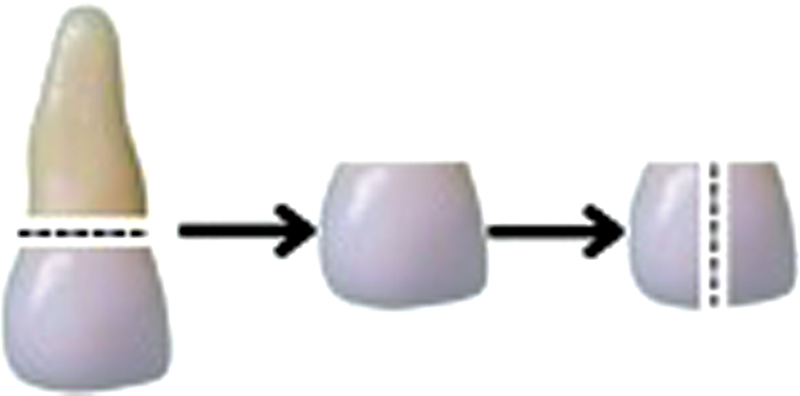
The specimens were prepared by precisely sectioning the crowns at the cementoenamel junction using a low-speed diamond saw with water cooling. This cutting was done in the mesiodistal direction, resulting in two specimens from each crown ([Fig. 1]). The exposed surfaces of the tooth sections were coated with acid-resistant nail varnish. These tooth slices were then preserved at 4°C until they were suitable for use, per ISO/TS 11405 recommendations.
Calcium Oxide Preparation
Fish bones were cut to pieces measuring approximately 2 to 5 mm. This step increases the surface area for subsequent processes. One kilogram of fish bone was boiled for 2 hours to eliminate the attached fish flesh and any other organic contaminants from the bones. After boiling, the bones were dried for 2 days. This drying period ensures that water and any remaining organic material from the boiling process are completely removed. Dried bone was mashed using a ball milling machine into a fine powder. Samples were then calcined using a furnace at 1,000°C for 5 hours.[22] [24] [36] The calcination process aimed to remove residual organic elements and noncalcium metals while breaking down calcium carbonate (CaCO3) within bones into calcium oxide (CaO). This CaO serves as a precursor to produce HAP powder.[17]
Synthesis of Hydroxyapatite
HAP was synthesized through the precipitation method. CaO powder derived from fish bones was mixed with HNO3 solution. This reaction yields a solution of calcium nitrate (Ca(NO3)2). Subsequently, this Ca(NO3)2 solution was mixed with phosphoric acid (H3PO4) solution while being heated to 40°C at a speed of 300 rpm. The pH of the resulting solution was initially measured and then adjusted to pH 10 by adding ammonium hydroxide (NH4OH). The process was conducted over a 24-hour period to generate a settled precipitate. This precipitate was subsequently filtered to separate HAP from the liquid and then rinsed with distilled water to eliminate byproducts, particularly ammonium nitrate (NH4NO3). The resulting precipitates were then dried in an oven at 110°C for 5 hours to remove any remaining moisture. These dry precipitates were re-sintered at a furnace at 900°C for 5 hours to enhance the crystallinity and stability of HAP. As a result, synthesized HAP was obtained in the form of a white powder.[36]
Hydroxyapatite Paste
The HAP paste was prepared by heating distilled water, followed by adding nipagin and sodium carboxymethyl cellulose (NaCMC), and stirring until a homogeneous mixture was obtained. Fine HAP powder weighing 15 g, previously characterized using X-ray diffraction (XRD), was moistened with glycerin. Menthol was dissolved in alcohol and, after that, combined with the HAP powder that had been immersed in glycerin and the hot distilled water mixture. Then, menthol was added to the HAP water and stirred until a homogeneous paste was formed.[25]
Characterization of the Quality of Hydroxyapatite Paste
Homogeneity Test
The homogeneity of the toothpaste was assessed by applying a thin layer of 0.1 g of the product onto a glass surface, which was subsequently covered with another glass object and visually examined. Testing was conducted every week for 3 weeks.[37]
pH Test
Determination of the pH of the paste was carried out with a calibrated pH meter. The pH meter was calibrated using a standard pH solution.[37]
Viscosity Test
The toothpaste samples were subjected to a viscosity test using a Brookfield viscometer with spindle no. 6 at a speed of 2.0 rpm. Testing was carried out every week for 3 weeks of storage.[37]
Characterization of Hydroxyapatite Paste
The HAP powder was characterized using XRD PANalytical Aeris. This analysis was conducted to identify the mineralogy of the sample powder and to assess the crystals' structure, orientation, and size. The XRD study revealed HAP formation, derived from calcium precursors from the disintegration of mackerel bones and phosphate precursors obtained from diammonium hydrogen phosphate.[18] The function group was investigated using Fourier transform infrared (FTIR) Spectrophotometer Thermo Scientific Nicolet iS10 at wavenumber 500 to 4,000/cm.
Preparation of Hydroxyapatite Paste
NaCMC was prepared by mixing it with distilled water and stirring until homogenous solution was achieved. The HAP powder, previously characterized using XRD, was blended with glycerin and water. This glycerin mixture was then slowly incorporated into the prepared NaCMC while stirring to ensure a homogenous mixture.
Lesion Formation and Remineralization
The samples were placed in a demineralizing solution with a pH of 5.2 containing phosphate acid. They were kept in this solution for 48 hours at 37°C and a relative humidity of 50% to induce lesion formation. Following the demineralization procedure, the specimens were assigned randomly to two groups. Specimens of the first group were applied with HAP paste (15% w/v) for 30 minutes (case group). The specimens were thoroughly rinsed with distilled water and placed in artificial saliva for 24 hours. This procedure was repeated for 7 days. Specimens of the control group were immersed in artificial saliva. Artificial saliva was refreshed daily in all groups.
Enamel Microhardness Test
Every specimen was securely embedded within an acrylic block, oriented with the enamel surface facing outward ([Fig. 2]). Approximately 150 µm of the enamel layer was sequentially removed to prepare the sample through polishing with 240, 400, 600, and 1,200 grit sandpaper under water cooling. The microhardness of the tooth surface was measured by subjecting each sample to a 100-g load for 10 seconds at two distinct locations using a Vickers microhardness tester (Wilson Instruments 402 MVD). The Vickers hardness number (VHN) was determined by calculating the load (expressed in kilogram-force) divided by the surface area (measured in square millimeters). This was done using the formula in equation 1[38]:

VHN = F/A = 1.8544F/d 2, (1)
where F represents the applied load in kilogram-force and A is the surface area in square millimeters.
After determining the VHN, the average microhardness of the two measurement locations was calculated and then reported as the enamel's microhardness. The data distribution was assessed using the Shapiro–Wilk test. The mean values of the case and control groups were compared using the independent t-test.
Results
The analysis of the HAP sample from C. striata fish bone using the XRD curve profile and HighScore Plus software revealed that the sample was pure HAP (100% HAP, no other compounds). The chemical formula of the HAP was Ca10(PO4)6(OH)2, and it had a hexagonal shape/crystal structure with dimensions of a = 9.439 Å, b = 9.439 Å, and c = 6.886 Å. The space group of the HAP was P63/m, with a space group number of 176 ([Fig. 3]).[21] [39]

The XRD analysis revealed distinct peaks on the curve, indicating a hexagonal crystal lattice, a characteristic feature of the HAP compound. These peaks correspond to specific crystal lattice planes: (002), (012), (112), (030), (202), (221), (222), (230), (213), (321), (140), and (004). The corresponding angles two theta for these peaks were the following: 25.856, 28.100, 32.155, 32.843, 34.019, 40.372, 46.625, 48.504, 49.426, 50.392, 51.168, and 53.161 degrees. The majority of the peak widths appeared to be broad rather than narrow. The examination revealed a crystallinity level of around 37.09%.[21] [39]
The FTIR spectroscopic investigation focused on the functional groups present in the HAP sample derived from C. striata fish bone ([Fig. 4]). The hydroxyl (OH–) and phosphate (PO4 3–) functional groups were observed in the samples. The results of FTIR analysis of C. striata fish bone samples were found in the IR region with a wavelength of 500 to 3,500/cm. [Fig. 4] shows the peak around wave number 3,399.53/cm, corresponding to the –NH functional group, namely amine. Wave 1,636.54/cm showed absorption for the carbonyl group C = O or amide I functional group. Carboxylic acids exhibit an absorption of OH groups in the 3,300 to 3,500/cm area, specifically at a wave number of 3,399.53/cm. Within the range of wave numbers 500 to 1,100/cm, there were phosphate ions detected at wave numbers 570.68, 611.52, 669.35, 873.55, and 1,030.78/cm.[40] [41]

The HAP paste had a pH value of 7.8, indicating that it is neutral. According to the pH guidelines established by the Indonesian National Standard (SNI), which ranges from 4.5 to 10, the paste preparation is within the safe range for teeth and mouth usage. Through spreadability tests conducted with various weights, it was determined that the application of the HAP paste exhibited excellent spreading ability. The spreadability of the HAP paste derived from C. striata fish bone was 6.25 to 7.25 cm ([Table 1]). The accepted range for good spreadability, defined by the SNI, is 5 to 7 cm.[42]
|
Load (g) |
Diameter (cm) |
|---|---|
|
50 |
6.25 |
|
100 |
6.60 |
|
150 |
6.90 |
|
200 |
7.25 |
The 3-day homogeneity test confirmed that the HAP paste was homogeneous. This indicates that all the components in the paste were thoroughly blended and had a smooth consistency without any air pockets, distinct particles, or uneven texture ([Table 2]).[42]
|
Testing day |
Homogeneity |
|---|---|
|
1 |
Homogenous |
|
2 |
Homogenous |
|
3 |
Homogenous |
[Table 3] presents the average microhardness value (±standard deviation) of the two groups. The independent t-tests showed significant statistical differences between the paired groups. The microhardness values of samples treated with the HAP paste were considerably more significant than those of the control group (p = 0.00).
|
Group |
N |
Mean ± SD |
|
|---|---|---|---|
|
Microhardness |
Case |
13 |
356.1923 ± 25.2186 |
|
Control |
13 |
269.6862 ± 22.9325 |
Discussion
Producing HAP from fish bones is a cost-effective method that can also yield pure HAP.[20] Scientists have developed a method to produce HAP from fish bones since they are easily accessible. This technique involves calcination,[20] [43] [44] precipitation,[18] [45] sol-gel,[46] [47] alkali hydrolysis,[23] and hydrothermal.[44] In Indonesia, mackerel and tuna are commonly utilized to produce HAP. The selection of fish bone variety is frequently influenced by the quantity and arrangement of the bones, which tend to detach easily from the flesh.[48]
Through precipitation, Anggresani et al successfully obtained HAP from mackerel bone.[45] Before precipitation occurred, the bones of mackerel fish underwent calcination at 800°C to produce CaO. The precipitation process involved the combination of Ca(OH)2 suspension as a calcium precursor and (NH4)2HPO4 as a phosphate precursor. The resulting mixture was then sintering at 900°C for 5 hours. Mutmainnah et al used a modified precipitation technique to produce HAP from tuna bones. Prior to utilization, tuna fish bones underwent calcination to generate CaO, as done in prior research. Phosphoric acid solution was used as a precursor for HAP production, followed by sintering the resulting precipitates.[49] In this study, HAP was synthesized from the bones of C. striata fish using the precipitation method. This study employed the methodology established by Devitasari et al to produce HAP from the bones of tilapia fish (Oreochromis niloticus).[36] The chemical precipitation approach is advantageous because of its simplicity and cost-effectiveness. Another notable advantage of this approach is the production of nontoxic byproducts.[50]
The XRD analysis was employed to ascertain and validate the crystalline phase and quantify HAP's crystallinity level. The XRD analysis revealed that not all HAP compounds exhibited crystalline structure, namely a hexagonal atomic arrangement. Some were still amorphous (i.e., the atomic arrangement was irregular). HAP had a crystallinity level of around 37.09%. This diverges from the research findings of Anggraini et al.[51] They employed the calcination technique to produce HAP from C. striata fish bone. Their findings demonstrated 59.5 and 88.9% crystallinity at 600 and 800°C, respectively.[46] Increasing the temperature during the calcination process leads to a greater formation of HAP phases. The peaks exhibit enhanced acuity with rising temperatures. The width of the line decreases, suggesting a higher degree of crystallinity.[52]
The FTIR analysis conducted in this study revealed the presence of specific functional groups in the materials, including amine (–NH), phosphate (PO4 2–), carbonate (CO3 2–), and hydroxyl (OH–) groups. Tarmidzi et al's statement supported the findings of this research, confirming that the wave number regions 1,610 to 1,690 and 625 to 767/cm are indeed part of the analysis results for globular protein compounds. Additionally, it was observed that albumin extract protein contains multiple –NH groups, which are amine groups. There were more amine groups than O-H groups or hydroxyl groups. The phosphate group exhibited distinct properties at specific wave numbers within the 500 to 1,100/cm range. These included wave numbers at 570.68, 611.52, 669.35, 873.55, and 1,030.78/cm.[40] [41]
The HAP paste was found to meet the requirements outlined in the SNI (12-3524-1995) based on the results of the characteristic tests, which included pH value, homogeneity, and spreadability. The pH of the HAP paste was classified as neutral, indicating that it is safe for application on the teeth and in the mouth. Pastes with excessively acidic pH levels might irritate the mouth cavity. A homogeneous paste preparation can produce maximum effect because the paste's ingredients have been mixed evenly.[53] To evaluate the application properties of the paste, a spreadability test was performed to measure its ability to spread equally upon contact with the sample. According to Warnida et al, the capacity to spread is crucial when formulating pastes. It has a crucial impact on the successful administration of active substances to the enamel at the appropriate dosage, ease of application, the force required to dispense it from the packaging, and its reception by customers.[54]
Microhardness testing, mainly using the Vickers harness technique, is a valuable tool in dentistry for evaluating the mechanical characteristics and surface alterations in enamel, such as demineralization and remineralization.[7] The enamel hardness test results demonstrated an increase in enamel hardness in the case group following the application of the HAP paste. The average enamel hardness value in the case group was 356.2 VHN, while the average enamel hardness value in the control group was 269.7 VHN. The two groups exhibited a notable disparity in enamel hardness, likely attributed to the remineralization capability of HAP derived from C. striata fish bones. The results of this research are comparable to the results of Devitasari et al's research, which stated that the HAP paste from tilapia bone waste can increase the surface hardness of enamel. An increase in enamel hardness occurred after the application of 10 and 15% tilapia fish bone HAP for 7 days.[36]
Dental enamel primarily comprises HAP, a mineral of calcium, and phosphate ions. Enamel is characterized by a highly organized structure, with HAP crystals forming tightly packed enamel rods that extend from the dentin–enamel junction to the outer surface of the tooth enamel. Interspersed between these enamel rods are areas referred to as enamel inner rods.[2] The highly organized three-dimensional structure of enamel is essential for its mechanical strength. It allows enamel to withstand the forces of biting and chewing, making it one of the hardest substances in the human body. The compact structure of enamel rods and their high mineral content make enamel resistant to microbial attacks and reflect lights.[55] Under normal circumstances, a dynamic equilibrium exists between the HAP minerals within the tooth's structure and oral fluids.[56] Demineralization, on the other hand, denotes the net reduction of calcium and phosphate ions from the tooth's structure. This occurs when acids produced by bacteria in dental plaque become active and dissolve HAP crystals. In contrast, remineralization reintroduces calcium and phosphate ions into the enamel structure. This restorative process occurs when supersaturated oral fluids, abundant in calcium and phosphate, come into contact with the previously demineralized enamel.[57]
Calcium and phosphate ions can be obtained by applying HAP on enamel surfaces, contributing to enamel remineralization.[58] HAP obtained from natural sources like plants, animals, biogenic materials, and aquatic sources offers an eco-friendly alternative to synthetic HAP. Natural HAP is inexpensive and abundant.[7] One source of HAP from natural materials is C. striata fish bone. Channa striata is endemic to waters throughout Indonesia, especially in wetland areas such as South Kalimantan. Channa striata fish bones have a high mineral content of calcium and phosphate. The calcium and phosphate content of C. striata fish bones can be used to make HAP paste to initiate remineralization.[22] According to the findings of Rosmawati et al, the bones of C. striata fish contain 21.74/900 to 1,000 g of calcium and 12.82/900 to 1,000 g of phosphorus.[35] Tawali et al's research findings indicate that the C. striata fish powder contains 35.85 mg/g of calcium and 0.70 mg/g of phosphorus minerals when reduced to nanosizes. The snakehead powder without nanosize contains 34.4 mg/g of calcium and 0.69 mg/g of phosphorus minerals.[26]
HAP acts as a reservoir of calcium and phosphate ions, essential for remineralizing tooth enamel and dentin. It can be incorporated into the porous tooth structure that results from the caries process. This incorporation helps replenish the lost minerals, increasing the mineral content and hardness of the affected tooth surfaces.[59] HAP plays a significant role in enhancing the degree of remineralization, particularly in acidic conditions, by increasing the supply of calcium and phosphorus ions to the demineralized zone.[60]
HAP contributes to maintaining an oversaturated state of enamel minerals. This oversaturation actively promotes the formation of a uniform apatite surface, thereby reducing the likelihood of demineralization and facilitating remineralization.[61] This effect may be attributed to depositing a fresh, uniform apatite surface layer on the previously demineralized area. This deposition aids in the restoration of lost minerals, ultimately boosting the mineral content and hardness of the affected tooth surfaces.[62] The newly formed apatite surface layer acts as a protective barrier against acid attacks and aids in restoring the enamel structure. HAP further promotes the buildup of minerals in the outer layer of caries lesions, creating a densely mineralized surface. This outer layer acts as a barrier, preventing mineral ions from infiltrating the deeper portions of the demineralized lesions, which could potentially halt the progression of dental decay.[60]
HAP infiltrates the pores formed by the breakdown of calcium and phosphate ions in early enamel lesions.[10] It efficiently replenishes the missing ions and forms a synthetic enamel layer on the tooth surface. This synthetic layer acts as a “sacrificial layer” during future acid attacks, providing a protective barrier that helps prevent further demineralization of the underlying natural enamel. In dentin, HAP penetrates the demineralized collagen matrix. This action serves as a scaffold for remineralization and provides a localized source of calcium and phosphate.[3]
HAP functions as a protective coating on the teeth's surface and a component of dental biofilms.[11] When acids from food or oral bacteria come into contact with the particulate HAP in the dental biofilm, it will be dissolved. This dissolution results in the release of calcium ions. The release of calcium ions increases the pH of the dental plaques. This elevated pH makes the acidic environment less favorable for further demineralization.[63]
HAP has a superior remineralization potential compared to fluoride, primarily due to its ability to penetrate deeper into the affected area. Fluoride remineralization primarily targets the surface layer of dental lesions. It works by promoting the redeposition of minerals, primarily fluoride, calcium, and phosphate ions, on the layer of the lesion.[64] The remineralization facilitated by HAP seems to extend deeper into the subsurface layer of the lesion and exhibit a more uniform distribution. HAP particles act as templates of the de novo formation of HAP crystals around each particle. This process continuously attracts large amounts of calcium and phosphate ions from the surrounding solution into the tooth tissue.[65] This biomimetic mineralization process promotes crystal integrity and growth and enables regeneration of the enamel and dentin.[63]
Conclusion
HAP was successfully synthesized from C. striata fish bones using the precipitation method. The HAP paste from C. striata fish bone stimulates the remineralization of primary teeth, characterized by an increase in enamel surface hardness. Therefore, the HAP paste has the potential to be applied as a caries preventive agent for primary teeth.
Conflict of Interest
None declared.
Acknowledgments
The authors acknowledge the Faculty of Dentistry, Universitas Lambung Mangkurat, and Advanced Characterization Laboratories Serpong, National Research and Innovation Institute, BRIN, for providing facilities and scientific and technical guidance.
-
References
- 1 Grohe B, Mittler S. Advanced non-fluoride approaches to dental enamel remineralization: the next level in enamel repair management. Biomater Biosyst 2021; 4: 100029
- 2 Shaik I, Dasari B, Shaik A. et al. Functional role of inorganic trace elements on enamel and dentin formation: a review. J Pharm Bioallied Sci 2021; 13 (6, Suppl 2): S952-S956
- 3 Chen L, Al-Bayatee S, Khurshid Z, Shavandi A, Brunton P, Ratnayake J. Hydroxyapatite in oral care products: a review. Materials (Basel) 2021; 14 (17) 4865
- 4 O'Hagan-Wong K, Enax J, Meyer F, Ganss B. The use of hydroxyapatite toothpaste to prevent dental caries. Odontology 2022; 110 (02) 223-230
- 5 Moradian-Oldak J. Protein-mediated enamel mineralization. Front Biosci (Landmark Ed) 2012; 17 (06) 1996-2023
- 6 Achmad H, Djais A, Hatta LI. et al. The impact of using fluoride in pediatric dentistry: a systematic review. Ann Rom Soc Cell Biol 2021; 25: 2816-2839
- 7 Mathirat A, Dalavi PA, Prabhu A. et al. Remineralizing potential of natural nano-hydroxyapatite obtained from Epinephelus chlorostigma in artificially induced early enamel lesion: an in vitro study. Nanomaterials (Basel) 2022; 12 (22) 3993
- 8 Enax J, Fabritius HO, Fabritius-Vilpoux K, Amaechi BT, Meyer F. Modes of action and clinical efficacy of particulate hydroxyapatite in preventive oral health care: state of the art. Open Dent J 2019; 13 (01) 274-287
- 9 Limeback H, Enax J, Meyer F. Biomimetic hydroxyapatite and caries prevention: a systematic review and meta-analysis. Can J Dent Hyg 2021; 55 (03) 148-159
- 10 Ionescu AC, Degli Esposti L, Iafisco M, Brambilla E. Dental tissue remineralization by bioactive calcium phosphate nanoparticles formulations. Sci Rep 2022; 12 (01) 5994
- 11 Sudradjat H, Meyer F, Loza K, Epple M, Enax J. In vivo effects of a hydroxyapatite-based oral care gel on the calcium and phosphorus levels of dental plaque. Eur J Dent 2020; 14 (02) 206-211
- 12 Khurshid Z, Alfarhan MFA, Bayan Y. et al. Development, physicochemical characterization and in-vitro biocompatibility study of dromedary camel dentine derived hydroxyapatite for bone repair. PeerJ 2023; 11: e15711
- 13 Khurshid Z, Alfarhan MF, Mazher J. et al. Extraction of hydroxyapatite from camel bone for bone tissue engineering application. Molecules 2022; 27 (22) 7946
- 14 Khurshid Z, Alqurashi H, Ashi H, Gen Dent EJ. Advancing environmental sustainability in dentistry and oral health. Eur J Gen Dent 2024; 13: 264-268
- 15 Grose J, Burns L, Mukonoweshuro R. et al. Developing sustainability in a dental practice through an action research approach. Br Dent J 2018; 225 (05) 409-413
- 16 Hossain MS, Uddin MN, Sarkar S, Ahmed S. Crystallographic dependency of waste cow bone, hydroxyapatite, and β-tricalcium phosphate for biomedical application. J Saudi Chem Soc 2022; 26 (06) 101559
- 17 Terzioğlu P, Öğüt H, Kalemtaş A. Natural calcium phosphates from fish bones and their potential biomedical applications. Mater Sci Eng C 2018; 91: 899-911
- 18 Anggresani L, Sari YN. , Rahmadevi. Hydroxyapatite (HAp) from tenggiri fish bones as abrasive material in toothpaste formula. J Kimia Valensi 2021; 7 (01) 1-9
- 19 Maulidah, Dwipura Hasbullah I, Uli Arta Panjaitan F. Biocompability test of Haruan fish (Channa striata) bone hydroxyapatite to fibroblast cell as periodontal pocket therapy (In vitro study on BHK-21 fibroblast cell with hydroxyapatite of Haruan fish bone (Channa striata) as bone graft material). Dentino J Kedokteran Gigi 2018; 3 (02) 150-155
- 20 Shi P, Liu M, Fan F, Yu C, Lu W, Du M. Characterization of natural hydroxyapatite originated from fish bone and its biocompatibility with osteoblasts. Mater Sci Eng C 2018; 90: 706-712
- 21 Surya P, Nithin A, Sundaramanickam A, Sathish M. Synthesis and characterization of nano-hydroxyapatite from Sardinella longiceps fish bone and its effects on human osteoblast bone cells. J Mech Behav Biomed Mater 2021; 119: 104501
- 22 Muryati M, Loekitowati Hariani P, Said M. Preparation and characterization nanoparticle calcium oxide from snakehead fish bone using ball milling method. Indones J Fundamental Appl Chem 2019; 4 (03) 111-115
- 23 Herpandi, Hanif I, Widiastuti I, Sudirman S. Hydroxyapatite characteristics from snakehead fish (Channa striata) bone via alkali treatment followed by calcination method. Trop J Nat Prod Res 2024; 8 (02) 6147-6151
- 24 Hariani PL, Muryati M, Said M, Salni S. Synthesis of nano-hydroxyapatite from snakehead (Channa striata) fish bone and its antibacterial properties. Key Eng Mater 2020; 840: 293-299
- 25 Dewi N, Rahmadella A, Hatta I, Apriasari ML, Putri KDT. Antibacterial activity of nano-hydroxyapatite paste of snakehead fish bone against S. mutans: an in vitro study. Padjadjaran J Dent 2024; 36 (01) 9-16
- 26 Tawali AB, Wakiah N, Qanitah K, Asfar M, Sukendar NK. , Metusalach. The effect of sonication time on physicochemical profiles of the nanocalsium from snake-head fish bone (Channa striata). IOP Conf Ser Earth Environ Sci 2019; 355 (01) 012091
- 27 Kusuma HH, Sifah L, Anggita SS. The characterization of hydroxyapatite from blood clam shells and eggs shells: synthesis by hydrothermal method. J Phys: Conf Ser 2021; 1918 (02) 022040
- 28 Wibisono Y, Ummah SR, Hermanto MB, Djoyowasito G, Noviyanto A. Slow-release hydroxyapatite fertilizer from crab shells waste for sustainable crop production. Results Eng 2024; 21: 101781
- 29 Suresh Kumar C, Dhanaraj K, Vimalathithan RM, Ilaiyaraja P, Suresh G. Hydroxyapatite for bone related applications derived from sea shell waste by simple precipitation method. J Asian Ceram Soc 2020; 8 (02) 416-429
- 30 Umesh M, Choudhury DD, Shanmugam S. et al. Eggshells biowaste for hydroxyapatite green synthesis using extract piper betel leaf: evaluation of antibacterial and antibiofilm activity. Environ Res 2021; 200: 111493
- 31 Ibrahim AR, Li X, Zhou Y. et al. Synthesis of spongy-like mesoporous hydroxyapatite from raw waste eggshells for enhanced dissolution of ibuprofen loaded via supercritical CO2 . Int J Mol Sci 2015; 16 (04) 7960-7975
- 32 Mousavi SM, Hashemi SA, Yousefi k. et al. Antibacterial and cytotoxic efficacy of nano-hydroxyapatite synthesized from eggshell and sheep bones bio waste. Research Square 2022;
- 33 dos Santos Horta MK, Westin C, da Rocha DN. et al. Hydroxyapatite from biowaste for biomedical applications: obtainment, characterization and in vitro assays. Mater Res 2023; 26: e20220466
- 34 Alviodinasyari R, Pribadi ES, Soejoedono RD. Soluble protein concentration in snakehead fish albumin Bogor origin (Channa striata and Channa micropeltes). J Vet 2019; 20 (03) 436
- 35 Rosmawati, Bakar Tawali A, Laga A. Karakteristik kimia tulang ikan gabus (Channa striata) dari bobot badan berbeda [Chemical characteristics of snakehead fish bones (Channa striata) of different body weights]. J Inovasi Sains Dan Teknologi (INSTEK) 2019; 2 (01) 63-80
- 36 Devitasari SP, Hudiyati M, Anastasia D. Effect of hydroxyapatite from waste of tilapia bone (Oreochromis niloticus) on the surface hardness of enamel. J Phys: Conf Ser 2019; 1246 (01) 012009
- 37 Abidin AZ. Formulasi pasta gigi ekstrak etanol daun kenikir dan uji aktivitas antibakteri terhadap Streptococcus mutans. [Formulation of toothpaste with ethanol extract of cosmos leaves and antibacterial activity test against Streptococcus mutans]. Farmakologika J Farmasi 2023; 19 (02) 138-152
- 38 Comba A, Scotti N, Maravić T. et al. Vickers hardness and shrinkage stress evaluation of low and high viscosity bulk-fill resin composite. Polymers (Basel) 2020; 12 (07) 1477
- 39 Acharya P, Kupendra M, Fasim A. et al. Synthesis of nano hydroxyapatite from Hypopthalmichthys molitrix (silver carp) bone waste by two different methods: a comparative biophysical and in vitro evaluation on osteoblast MG63 cell lines. Biotechnol Lett 2022; 44 (10) 1175-1188
- 40 Indrani DJ, Lukitowati F, Yulizar Y. Preparation of chitosan/collagen blend membranes for wound dressing: a study on FTIR spectroscopy and mechanical properties. IOP Conf Ser Mater Sci Eng 2017; 202 (01) 12020
- 41 Tarmidzi FM, Tarihoran CRU, Jarkasih FR. Formulasi dan evaluasi karakteristik salep herbal dengan ekstrak binahong (Andradera cordifolia) dan ikan gabus (Channa striata) [Formulation and evaluation of herbal ointment containing extract of binahong (Andradera cordifolia) and catfish (Channa striata)]. Paper presented at: 2nd Seminastika; October 16-17, 2019 ; Balikpapan, Indonesia
- 42 Mahdalin A, Widarsih E, Harismah DK. Pengujian sifat fisika dan sifat kimia formulasi pasta gigi gambir dengan pemanis alami daun stevia [Physical and chemical properties of Uncaria toothpaste formulation with natural sweetener stevia leaves]. URECOL. 2017. Accessed October 13, 2023 at: https://journal.unimma.ac.id/index.php/urecol/article/view/1250
- 43 Venkatesan J, Qian ZJ, Ryu B, Thomas NV, Kim SK. A comparative study of thermal calcination and an alkaline hydrolysis method in the isolation of hydroxyapatite from Thunnus obesus bone. Biomed Mater 2011; 6 (03) 035003
- 44 Tri N, Trang TND, Trinh NHD. et al. Hydrothermal and calcination regimes and characteristics of nanohydroxyapatite synthesized from salmon bones. J Biochem Technol 2020; 11 (02) 82-87
- 45 Anggresani L, Perawati S, Dianal F, Sutrisno D. Pengaruh Variasi Perbandingan Mol Ca/P Dalam Pembuatan Hidroksiapatit dari Tulang Ikan Tenggiri (Scomberomorus guttatus). J Farmasi Higea 2020; 12 (01) 55-64
- 46 Esmael SK, Jassim RK, Mahdi R. Preparation and characterization of nano-hydroxyapatite particles and chitosan by sol-gel method (in vitro study). Indian J Forensic Med Toxicol 2020; 14 (02) 593-599
- 47 Rahmayuni Zein U, Anggresani L, Yulianis Y. Pengaruh waktu sintering terhadap hidroksiapatit berpori tulang ikan tenggiri dengan proses sol-gel [Effect of sintering time on porous hydroxyapatite from mackerel fish bone using sol-gel process]. Chempublish J 2020; 5 (01) 46-56
- 48 Afriani F. , Siswoyo, Amelia R, Hudatwi M. , Zaitun, Tiandho Y. Hydroxyapatite from natural sources: methods and its characteristics. IOP Conf Ser Earth Environ Sci 2020; 599 (01) 012055
- 49 Mutmainnah M, Chadijah S, Rustiah WO. Hidroksiapatit dari tulang ikan tuna sirip kuning (Thunnus albacares) dengan metode presipitasi [Hydroxyapatite from yellowfin tuna (Thunnus albacares) bones by precipitation method]. Al-Kimia 2017; 5 (02) 119-126
- 50 Yelten-Yilmaz A, Yilmaz S. Wet chemical precipitation synthesis of hydroxyapatite (HA) powders. Ceram Int 2018; 44 (08) 9703-9710
- 51 Mutia AnggrainiR, Restianingsih T, Deswardani F, Fendriani Y, Ananda R, Purba P. Characterization of hydroxyapatite from Channa striata and Scomberomorus commerson fish bone by heat treatment. Journal Online of Physics 2023; 9 (01) 49-54
- 52 Ahmed YMZ, El-Sheikh SM, Zaki ZI. Changes in hydroxyapatite powder properties via heat treatment. Bull Mater Sci 2015; 38 (07) 1807-1819
- 53 Gusnawati, Sabara Z. , Munira, Bakhri S. Karakterisasi mutu pasta gigi dengan penambahan garam dan virgin coconut oil (VCO) ditinjau dari SNI 12–3524–1995 [Characterization of the quality of toothpaste with the addition of salt and virgin coconut oil (VCO) reviewed from SNI 12–3524–1995]. J Industri Hasil Perkebunan 2022; 17 (01) 41-49
- 54 Warnida H, Juliannor A, Sukawaty Y. Formulasi Pasta gigi gel ekstrak etanol bawang dayak (Eleutherine bulbosa (Mill.) Urb.) [Toothpaste gel formulation of Dayak onion ethanol extract (Eleutherine bulbosa (Mill.) Urb.)]. J Sains Farmasi Klinis 2016; 3 (01) 42
- 55 Tosco V, Vitiello F, Monterubbianesi R. et al. Assessment of the remineralizing potential of biomimetic materials on early artificial caries lesions after 28 days: an in vitro study. Bioengineering (Basel) 2023; 10 (04) 462
- 56 Anil AI, Ibraheem WA, Meshni A, Preethanath R, Anil S. Demineralization and remineralization dynamics and dental caries. In: Rusu L-C, Cosmina Ardelean L. eds. Dental Caries: The Selection of Restoration Methods and Restorative Materials. London: InTechOpen Limited; 2022
- 57 Carey CM. Remineralization of early enamel lesions with apatite-forming salt. Dent J 2023; 11 (08) 182
- 58 Febriani M, Amelia H, Alawiyah T, Rachmawati E. The potential of hydroxyapatite toothpaste towards the hypersensitive tooth. Int J Med Sci Clin Invent 2021; 8 (12) 5849-5857
- 59 Pushpalatha C, Gayathri VS, Sowmya SV. et al. Nanohydroxyapatite in dentistry: a comprehensive review. Saudi Dent J 2023; 35 (06) 741-752
- 60 Anil A, Ibraheem WI, Meshni AA, Preethanath RS, Anil S. Nano-hydroxyapatite (nHAp) in the remineralization of early dental caries: a scoping review. Int J Environ Res Public Health 2022; 19 (09) 5629
- 61 Huang S, Gao S, Cheng L, Yu H. Combined effects of nano-hydroxyapatite and Galla chinensis on remineralisation of initial enamel lesion in vitro. J Dent 2010; 38 (10) 811-819
- 62 Juntavee A, Juntavee N, Hirunmoon P. Remineralization potential of nanohydroxyapatite toothpaste compared with tricalcium phosphate and fluoride toothpaste on artificial carious lesions. Int J Dent 2021; 2021: 5588832
- 63 Meyer F, Enax J, Amaechi BT. et al. Hydroxyapatite as remineralization agent for children's dental care. Front Dent Med 2022; 3: 1-10
- 64 Amaechi BT, Phillips TS, Evans V. et al. The potential of hydroxyapatite toothpaste to prevent root caries: a pH-cycling study. Clin Cosmet Investig Dent 2021; 13: 315-324
- 65 Amaechi BT, van Loveren C. Fluorides and non-fluoride remineralization systems. Monogr Oral Sci 2013; 23: 15-26
Address for correspondence
Publication History
Article published online:
08 November 2024
© 2024. The Author(s). This is an open access article published by Thieme under the terms of the Creative Commons Attribution License, permitting unrestricted use, distribution, and reproduction so long as the original work is properly cited. (https://creativecommons.org/licenses/by/4.0/)
Thieme Medical and Scientific Publishers Pvt. Ltd.
A-12, 2nd Floor, Sector 2, Noida-201301 UP, India
-
References
- 1 Grohe B, Mittler S. Advanced non-fluoride approaches to dental enamel remineralization: the next level in enamel repair management. Biomater Biosyst 2021; 4: 100029
- 2 Shaik I, Dasari B, Shaik A. et al. Functional role of inorganic trace elements on enamel and dentin formation: a review. J Pharm Bioallied Sci 2021; 13 (6, Suppl 2): S952-S956
- 3 Chen L, Al-Bayatee S, Khurshid Z, Shavandi A, Brunton P, Ratnayake J. Hydroxyapatite in oral care products: a review. Materials (Basel) 2021; 14 (17) 4865
- 4 O'Hagan-Wong K, Enax J, Meyer F, Ganss B. The use of hydroxyapatite toothpaste to prevent dental caries. Odontology 2022; 110 (02) 223-230
- 5 Moradian-Oldak J. Protein-mediated enamel mineralization. Front Biosci (Landmark Ed) 2012; 17 (06) 1996-2023
- 6 Achmad H, Djais A, Hatta LI. et al. The impact of using fluoride in pediatric dentistry: a systematic review. Ann Rom Soc Cell Biol 2021; 25: 2816-2839
- 7 Mathirat A, Dalavi PA, Prabhu A. et al. Remineralizing potential of natural nano-hydroxyapatite obtained from Epinephelus chlorostigma in artificially induced early enamel lesion: an in vitro study. Nanomaterials (Basel) 2022; 12 (22) 3993
- 8 Enax J, Fabritius HO, Fabritius-Vilpoux K, Amaechi BT, Meyer F. Modes of action and clinical efficacy of particulate hydroxyapatite in preventive oral health care: state of the art. Open Dent J 2019; 13 (01) 274-287
- 9 Limeback H, Enax J, Meyer F. Biomimetic hydroxyapatite and caries prevention: a systematic review and meta-analysis. Can J Dent Hyg 2021; 55 (03) 148-159
- 10 Ionescu AC, Degli Esposti L, Iafisco M, Brambilla E. Dental tissue remineralization by bioactive calcium phosphate nanoparticles formulations. Sci Rep 2022; 12 (01) 5994
- 11 Sudradjat H, Meyer F, Loza K, Epple M, Enax J. In vivo effects of a hydroxyapatite-based oral care gel on the calcium and phosphorus levels of dental plaque. Eur J Dent 2020; 14 (02) 206-211
- 12 Khurshid Z, Alfarhan MFA, Bayan Y. et al. Development, physicochemical characterization and in-vitro biocompatibility study of dromedary camel dentine derived hydroxyapatite for bone repair. PeerJ 2023; 11: e15711
- 13 Khurshid Z, Alfarhan MF, Mazher J. et al. Extraction of hydroxyapatite from camel bone for bone tissue engineering application. Molecules 2022; 27 (22) 7946
- 14 Khurshid Z, Alqurashi H, Ashi H, Gen Dent EJ. Advancing environmental sustainability in dentistry and oral health. Eur J Gen Dent 2024; 13: 264-268
- 15 Grose J, Burns L, Mukonoweshuro R. et al. Developing sustainability in a dental practice through an action research approach. Br Dent J 2018; 225 (05) 409-413
- 16 Hossain MS, Uddin MN, Sarkar S, Ahmed S. Crystallographic dependency of waste cow bone, hydroxyapatite, and β-tricalcium phosphate for biomedical application. J Saudi Chem Soc 2022; 26 (06) 101559
- 17 Terzioğlu P, Öğüt H, Kalemtaş A. Natural calcium phosphates from fish bones and their potential biomedical applications. Mater Sci Eng C 2018; 91: 899-911
- 18 Anggresani L, Sari YN. , Rahmadevi. Hydroxyapatite (HAp) from tenggiri fish bones as abrasive material in toothpaste formula. J Kimia Valensi 2021; 7 (01) 1-9
- 19 Maulidah, Dwipura Hasbullah I, Uli Arta Panjaitan F. Biocompability test of Haruan fish (Channa striata) bone hydroxyapatite to fibroblast cell as periodontal pocket therapy (In vitro study on BHK-21 fibroblast cell with hydroxyapatite of Haruan fish bone (Channa striata) as bone graft material). Dentino J Kedokteran Gigi 2018; 3 (02) 150-155
- 20 Shi P, Liu M, Fan F, Yu C, Lu W, Du M. Characterization of natural hydroxyapatite originated from fish bone and its biocompatibility with osteoblasts. Mater Sci Eng C 2018; 90: 706-712
- 21 Surya P, Nithin A, Sundaramanickam A, Sathish M. Synthesis and characterization of nano-hydroxyapatite from Sardinella longiceps fish bone and its effects on human osteoblast bone cells. J Mech Behav Biomed Mater 2021; 119: 104501
- 22 Muryati M, Loekitowati Hariani P, Said M. Preparation and characterization nanoparticle calcium oxide from snakehead fish bone using ball milling method. Indones J Fundamental Appl Chem 2019; 4 (03) 111-115
- 23 Herpandi, Hanif I, Widiastuti I, Sudirman S. Hydroxyapatite characteristics from snakehead fish (Channa striata) bone via alkali treatment followed by calcination method. Trop J Nat Prod Res 2024; 8 (02) 6147-6151
- 24 Hariani PL, Muryati M, Said M, Salni S. Synthesis of nano-hydroxyapatite from snakehead (Channa striata) fish bone and its antibacterial properties. Key Eng Mater 2020; 840: 293-299
- 25 Dewi N, Rahmadella A, Hatta I, Apriasari ML, Putri KDT. Antibacterial activity of nano-hydroxyapatite paste of snakehead fish bone against S. mutans: an in vitro study. Padjadjaran J Dent 2024; 36 (01) 9-16
- 26 Tawali AB, Wakiah N, Qanitah K, Asfar M, Sukendar NK. , Metusalach. The effect of sonication time on physicochemical profiles of the nanocalsium from snake-head fish bone (Channa striata). IOP Conf Ser Earth Environ Sci 2019; 355 (01) 012091
- 27 Kusuma HH, Sifah L, Anggita SS. The characterization of hydroxyapatite from blood clam shells and eggs shells: synthesis by hydrothermal method. J Phys: Conf Ser 2021; 1918 (02) 022040
- 28 Wibisono Y, Ummah SR, Hermanto MB, Djoyowasito G, Noviyanto A. Slow-release hydroxyapatite fertilizer from crab shells waste for sustainable crop production. Results Eng 2024; 21: 101781
- 29 Suresh Kumar C, Dhanaraj K, Vimalathithan RM, Ilaiyaraja P, Suresh G. Hydroxyapatite for bone related applications derived from sea shell waste by simple precipitation method. J Asian Ceram Soc 2020; 8 (02) 416-429
- 30 Umesh M, Choudhury DD, Shanmugam S. et al. Eggshells biowaste for hydroxyapatite green synthesis using extract piper betel leaf: evaluation of antibacterial and antibiofilm activity. Environ Res 2021; 200: 111493
- 31 Ibrahim AR, Li X, Zhou Y. et al. Synthesis of spongy-like mesoporous hydroxyapatite from raw waste eggshells for enhanced dissolution of ibuprofen loaded via supercritical CO2 . Int J Mol Sci 2015; 16 (04) 7960-7975
- 32 Mousavi SM, Hashemi SA, Yousefi k. et al. Antibacterial and cytotoxic efficacy of nano-hydroxyapatite synthesized from eggshell and sheep bones bio waste. Research Square 2022;
- 33 dos Santos Horta MK, Westin C, da Rocha DN. et al. Hydroxyapatite from biowaste for biomedical applications: obtainment, characterization and in vitro assays. Mater Res 2023; 26: e20220466
- 34 Alviodinasyari R, Pribadi ES, Soejoedono RD. Soluble protein concentration in snakehead fish albumin Bogor origin (Channa striata and Channa micropeltes). J Vet 2019; 20 (03) 436
- 35 Rosmawati, Bakar Tawali A, Laga A. Karakteristik kimia tulang ikan gabus (Channa striata) dari bobot badan berbeda [Chemical characteristics of snakehead fish bones (Channa striata) of different body weights]. J Inovasi Sains Dan Teknologi (INSTEK) 2019; 2 (01) 63-80
- 36 Devitasari SP, Hudiyati M, Anastasia D. Effect of hydroxyapatite from waste of tilapia bone (Oreochromis niloticus) on the surface hardness of enamel. J Phys: Conf Ser 2019; 1246 (01) 012009
- 37 Abidin AZ. Formulasi pasta gigi ekstrak etanol daun kenikir dan uji aktivitas antibakteri terhadap Streptococcus mutans. [Formulation of toothpaste with ethanol extract of cosmos leaves and antibacterial activity test against Streptococcus mutans]. Farmakologika J Farmasi 2023; 19 (02) 138-152
- 38 Comba A, Scotti N, Maravić T. et al. Vickers hardness and shrinkage stress evaluation of low and high viscosity bulk-fill resin composite. Polymers (Basel) 2020; 12 (07) 1477
- 39 Acharya P, Kupendra M, Fasim A. et al. Synthesis of nano hydroxyapatite from Hypopthalmichthys molitrix (silver carp) bone waste by two different methods: a comparative biophysical and in vitro evaluation on osteoblast MG63 cell lines. Biotechnol Lett 2022; 44 (10) 1175-1188
- 40 Indrani DJ, Lukitowati F, Yulizar Y. Preparation of chitosan/collagen blend membranes for wound dressing: a study on FTIR spectroscopy and mechanical properties. IOP Conf Ser Mater Sci Eng 2017; 202 (01) 12020
- 41 Tarmidzi FM, Tarihoran CRU, Jarkasih FR. Formulasi dan evaluasi karakteristik salep herbal dengan ekstrak binahong (Andradera cordifolia) dan ikan gabus (Channa striata) [Formulation and evaluation of herbal ointment containing extract of binahong (Andradera cordifolia) and catfish (Channa striata)]. Paper presented at: 2nd Seminastika; October 16-17, 2019 ; Balikpapan, Indonesia
- 42 Mahdalin A, Widarsih E, Harismah DK. Pengujian sifat fisika dan sifat kimia formulasi pasta gigi gambir dengan pemanis alami daun stevia [Physical and chemical properties of Uncaria toothpaste formulation with natural sweetener stevia leaves]. URECOL. 2017. Accessed October 13, 2023 at: https://journal.unimma.ac.id/index.php/urecol/article/view/1250
- 43 Venkatesan J, Qian ZJ, Ryu B, Thomas NV, Kim SK. A comparative study of thermal calcination and an alkaline hydrolysis method in the isolation of hydroxyapatite from Thunnus obesus bone. Biomed Mater 2011; 6 (03) 035003
- 44 Tri N, Trang TND, Trinh NHD. et al. Hydrothermal and calcination regimes and characteristics of nanohydroxyapatite synthesized from salmon bones. J Biochem Technol 2020; 11 (02) 82-87
- 45 Anggresani L, Perawati S, Dianal F, Sutrisno D. Pengaruh Variasi Perbandingan Mol Ca/P Dalam Pembuatan Hidroksiapatit dari Tulang Ikan Tenggiri (Scomberomorus guttatus). J Farmasi Higea 2020; 12 (01) 55-64
- 46 Esmael SK, Jassim RK, Mahdi R. Preparation and characterization of nano-hydroxyapatite particles and chitosan by sol-gel method (in vitro study). Indian J Forensic Med Toxicol 2020; 14 (02) 593-599
- 47 Rahmayuni Zein U, Anggresani L, Yulianis Y. Pengaruh waktu sintering terhadap hidroksiapatit berpori tulang ikan tenggiri dengan proses sol-gel [Effect of sintering time on porous hydroxyapatite from mackerel fish bone using sol-gel process]. Chempublish J 2020; 5 (01) 46-56
- 48 Afriani F. , Siswoyo, Amelia R, Hudatwi M. , Zaitun, Tiandho Y. Hydroxyapatite from natural sources: methods and its characteristics. IOP Conf Ser Earth Environ Sci 2020; 599 (01) 012055
- 49 Mutmainnah M, Chadijah S, Rustiah WO. Hidroksiapatit dari tulang ikan tuna sirip kuning (Thunnus albacares) dengan metode presipitasi [Hydroxyapatite from yellowfin tuna (Thunnus albacares) bones by precipitation method]. Al-Kimia 2017; 5 (02) 119-126
- 50 Yelten-Yilmaz A, Yilmaz S. Wet chemical precipitation synthesis of hydroxyapatite (HA) powders. Ceram Int 2018; 44 (08) 9703-9710
- 51 Mutia AnggrainiR, Restianingsih T, Deswardani F, Fendriani Y, Ananda R, Purba P. Characterization of hydroxyapatite from Channa striata and Scomberomorus commerson fish bone by heat treatment. Journal Online of Physics 2023; 9 (01) 49-54
- 52 Ahmed YMZ, El-Sheikh SM, Zaki ZI. Changes in hydroxyapatite powder properties via heat treatment. Bull Mater Sci 2015; 38 (07) 1807-1819
- 53 Gusnawati, Sabara Z. , Munira, Bakhri S. Karakterisasi mutu pasta gigi dengan penambahan garam dan virgin coconut oil (VCO) ditinjau dari SNI 12–3524–1995 [Characterization of the quality of toothpaste with the addition of salt and virgin coconut oil (VCO) reviewed from SNI 12–3524–1995]. J Industri Hasil Perkebunan 2022; 17 (01) 41-49
- 54 Warnida H, Juliannor A, Sukawaty Y. Formulasi Pasta gigi gel ekstrak etanol bawang dayak (Eleutherine bulbosa (Mill.) Urb.) [Toothpaste gel formulation of Dayak onion ethanol extract (Eleutherine bulbosa (Mill.) Urb.)]. J Sains Farmasi Klinis 2016; 3 (01) 42
- 55 Tosco V, Vitiello F, Monterubbianesi R. et al. Assessment of the remineralizing potential of biomimetic materials on early artificial caries lesions after 28 days: an in vitro study. Bioengineering (Basel) 2023; 10 (04) 462
- 56 Anil AI, Ibraheem WA, Meshni A, Preethanath R, Anil S. Demineralization and remineralization dynamics and dental caries. In: Rusu L-C, Cosmina Ardelean L. eds. Dental Caries: The Selection of Restoration Methods and Restorative Materials. London: InTechOpen Limited; 2022
- 57 Carey CM. Remineralization of early enamel lesions with apatite-forming salt. Dent J 2023; 11 (08) 182
- 58 Febriani M, Amelia H, Alawiyah T, Rachmawati E. The potential of hydroxyapatite toothpaste towards the hypersensitive tooth. Int J Med Sci Clin Invent 2021; 8 (12) 5849-5857
- 59 Pushpalatha C, Gayathri VS, Sowmya SV. et al. Nanohydroxyapatite in dentistry: a comprehensive review. Saudi Dent J 2023; 35 (06) 741-752
- 60 Anil A, Ibraheem WI, Meshni AA, Preethanath RS, Anil S. Nano-hydroxyapatite (nHAp) in the remineralization of early dental caries: a scoping review. Int J Environ Res Public Health 2022; 19 (09) 5629
- 61 Huang S, Gao S, Cheng L, Yu H. Combined effects of nano-hydroxyapatite and Galla chinensis on remineralisation of initial enamel lesion in vitro. J Dent 2010; 38 (10) 811-819
- 62 Juntavee A, Juntavee N, Hirunmoon P. Remineralization potential of nanohydroxyapatite toothpaste compared with tricalcium phosphate and fluoride toothpaste on artificial carious lesions. Int J Dent 2021; 2021: 5588832
- 63 Meyer F, Enax J, Amaechi BT. et al. Hydroxyapatite as remineralization agent for children's dental care. Front Dent Med 2022; 3: 1-10
- 64 Amaechi BT, Phillips TS, Evans V. et al. The potential of hydroxyapatite toothpaste to prevent root caries: a pH-cycling study. Clin Cosmet Investig Dent 2021; 13: 315-324
- 65 Amaechi BT, van Loveren C. Fluorides and non-fluoride remineralization systems. Monogr Oral Sci 2013; 23: 15-26