Keywords
intracerebral hemorrhage - sleep - risk factor - chronic kidney disease - diabetes
Introduction
There have been numerous studies on the frequency and risk factors of ischemic stroke
that occur during sleep, commonly referred to as wake-up stroke.[1] Moreover, in recent years, mechanical thrombectomy has been actively performed in
eligible cases with wake-up stroke.[2] In contrast, studies on hemorrhagic stroke occurring during sleep remain exceedingly
scarce.[3]
[4] We conducted a study to identify the clinical characteristics and risk factors of
patients with spontaneous intracerebral hemorrhage (sICH) occurring during sleep.
Materials and Methods
Patient Selection
This was a single-center, retrospective study with a study period between January
2012 and December 2017. For all the sICH patients admitted via our emergency department
(ED), stroke types and presenting symptoms as well as detailed data on when, where,
and how their symptoms developed were registered on the database by ED residents.
Based on behaviors at the time of onset, sICH patients were classified into two groups:
those with onset during sleep (sleep group) and those with onset while awake (awake
group). For comparison, the proportion of the awake group was also searched for patients
with cerebral infarction (CI) and aneurysmal subarachnoid hemorrhage (SAH) treated
at our institution during the same period. Patients with ICH secondary to trauma,
vascular malformations (such as cerebral aneurysms and arteriovenous malformations),
abnormal vasculature (such as moyamoya disease), or infections (such as bacterial
endocarditis) were excluded.
Two-Group Comparison
Between the sleep group and the awake group, demographic variables including age,
sex, systolic blood pressure (BP) measured at ED, comorbidities (hypertension, history
of ischemic heart disease, history of stroke, diabetes mellitus, hyperlipidemia, chronic
kidney disease, smoking history, use of oral antiplatelet agents, and use of oral
anticoagulants), the Glasgow Coma Scale (GCS) score at admission, and the anatomical
location of the sICH (basal ganglia/subcortical/posterior fossa) were compared. The
GCS scores of 15, ≤8, and 3 were defined as indicators of alertness (GCS 15), coma
(GCS ≤8), and deep coma (GCS 3). Additionally, the outcomes at hospital discharge
evaluated with the modified Rankin scale (mRS) were compared: those with mRS scores
≤ were defined as having favorable outcomes. The National Institutes of Health Stroke
Scale (NIHSS) score was not analyzed in this study due to insufficient data collection
during the study period.
Multivariate Logistic Regression Analysis
A multivariate logistic regression analysis was conducted using the aforementioned
11 demographic variables (age, sex, hypertension, history of ischemic heart disease,
history of stroke, diabetes, hyperlipidemia, chronic kidney disease, smoking history,
use of oral antiplatelet agents, and use of oral anticoagulants) to identify potential
risk factors associated with sICH occurring during sleep.
Subgroup Analysis Based on the Mode of Clinical Presentations
The sleep group was further subdivided into three categories based on the mode of
presentation to the ED: (1) deficits noticed after waking up (deficits), (2) sudden
headache waking up patients (sudden headache), and (3) failure to wake up. Their proportions
and association with the outcomes were evaluated.
Ethical Approval
This study was approved by the institutional review board of the institution (approval
number: 17-0013; approval date: October 17, 2017) and conducted in accordance with
the Declaration of Helsinki. The need for informed consent from each participant was
waived by the institutional review board.
Statistical Analysis
Statistical analysis was performed using SPSS version 18.0 (SPSS Inc., Chicago, IL,
United States). For continuous variables (expressed as mean ± standard deviation),
Student's t-test was used, and Fisher's exact test was performed for categorical variables. A
p-value of less than 0.05 was considered statistically significant.
Results
Frequency
During the study period, a total of 710 ICH patients were brought to our ED. After
excluding 41 patients who met the exclusion criteria, 669 sICH patients were identified,
among whom 149 patients had unclear behavioral patterns at onset and excluded from
analysis. Ultimately, 520 sICH patients with a clear behavioral pattern at onset were
included for analysis. Among those, 119 patients (22.9%) had onset during sleep. During
the same period, 1,527 patients with CI and 257 patients with aneurysmal SAH were
brought to our ED, with sleep-onset CI occurring in 406 (26.7%) patients and sleep-onset
SAH occurring in 41 (16.0%) among those two groups. The proportion of the sleep group
among the sICH patients was lower than that of CI patients (26.7%) but was higher
than that of SAH patients (16.0%). A significant difference was noted between sICH
and SAH patients (p = 0.03; [Fig. 1]).
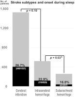 Fig. 1 The total number of acute stroke patients treated from 2011 to 2016 was as follows:
1,527 cases of cerebral infarctions, 520 cases of intracerebral hemorrhages (ICHs),
and 257 cases of subarachnoid hemorrhages (SAHs). Among these, the number of patients
with onset during sleep was 406 (cerebral infarctions), 119 (ICHs), and 41 (SAHs).
The proportion of ICHs with onset during sleep was significantly higher than that
SAHs (p = 0.03*).
Fig. 1 The total number of acute stroke patients treated from 2011 to 2016 was as follows:
1,527 cases of cerebral infarctions, 520 cases of intracerebral hemorrhages (ICHs),
and 257 cases of subarachnoid hemorrhages (SAHs). Among these, the number of patients
with onset during sleep was 406 (cerebral infarctions), 119 (ICHs), and 41 (SAHs).
The proportion of ICHs with onset during sleep was significantly higher than that
SAHs (p = 0.03*).
Two-Group Comparison
A comparison of demographic variables between the sleep group and the awake group
is shown in [Table 1]. No significant differences were observed between the two groups in terms of age
and gender ratio. While the sleep group exhibited lower systolic BP at ED arrival,
the difference was not statistically significant (179.9 ± 38.8 vs. 187.4 ± 40.0 mm
Hg; p = 0.07). Among the comorbidities, significant differences had been found for diabetes
(27.7% in the sleep group vs. 18.7% in the awake group; p = 0.04) and chronic kidney disease (24.4% in the sleep group vs. 12.7% in the awake
group; p = 0.004; [Table 1]). Regarding the neurological severity, no significant differences had been observed
between the two groups in terms of a GCS score of 15 (alert) or a GCS score of ≤8
(coma). However, the frequency of a GCS score of 3 (deep coma) was significantly higher
in the sleep group (14.3 vs. 6.7%; p = 0.01). In terms of the anatomical location of the hematoma, the sleep group tended
to have a higher frequency of basal ganglia hemorrhage and a lower frequency of subcortical
hemorrhage. However, the difference did not reach statistical significance (both p = 0.07).
Table 1
Comparison of demographic variables in sICH occurring during sleep and sICH occurring
during awake activities
|
Sleep (n = 119)
|
Awake (n = 401)
|
p-Value
|
|
Age (y)
|
66.7 ± 15.1
|
66.3 ± 12.7
|
0.80
|
|
Male:female
|
75:44
|
236:165
|
0.47
|
|
SBP at emergency department
|
179.9 ± 38.8
|
187.4 ± 40.0
|
0.07
|
|
Hypertension
|
88 (54.4%)
|
276 (44.2%)
|
0.31
|
|
Ischemic heart diseases
|
20 (16.8%)
|
59 (14.7%)
|
0.47
|
|
Prior stroke
|
34 (28.6%)
|
82 (20.4%)
|
0.08
|
|
Diabetes mellitus
|
33 (27.7%)
|
75 (18.7%)
|
0.04[a]
|
|
Hyperlipidemia
|
21 (17.6%)
|
61 (15.2%)
|
0.67
|
|
Chronic kidney diseases
|
29 (24.4%)
|
51 (12.7%)
|
0.004[a]
|
|
Smoking
|
28 (23.5%)
|
110 (27.4%)
|
0.48
|
|
Antiplatelet
|
27 (22.7%)
|
63 (15.7%)
|
0.10
|
|
Anticoagulant (warfarin 33, DOAC 7)
|
13 (10.9%)
|
27 (6.7%)
|
0.17
|
|
GCS 15
|
32 (26.9%)
|
119 (29.7%)
|
0.65
|
|
GCS ≤ 8
|
43 (36.1%)
|
118 (29.4%)
|
0.18
|
|
GCS 3
|
17 (14.3%)
|
27 (6.7%)
|
0.01[a]
|
|
Basal ganglia
|
81 (68.1%)
|
234 (58.3%)
|
0.07
|
|
Subcortex
|
14 (11.8%)
|
76 (19.0%)
|
0.07
|
|
Posterior fossa
|
25 (21.0%)
|
88 (21.9%)
|
0.90
|
Abbreviations: DOAC, direct oral anti-coagulant; GCS, Glasgow Coma Scale; SBP, systolic
blood pressure.
a Statistically significant.
Outcomes
A comparison of outcomes at hospital discharge between the two groups is shown in
[Table 2]. While no significant difference was observed between the two groups in the proportion
of favorable outcomes (13.4 vs. 14.7%; p = 0.88), the in-hospital mortality rate was significantly higher in the sleep group
(39.5 vs. 24.2%; p = 0.002).
Table 2
Comparison of outcomes evaluated at hospital discharge between sICH occurring during
sleep and sICH occurring during awake activities
|
Sleep (n = 119)
|
Awake (n = 401)
|
p-Value
|
|
Surgical intervention
|
6 (5.0%)
|
35 (8.7%)
|
0.25
|
|
Hospital stay (d)
|
25.8 ± 15.6
|
24.2 ± 14.0
|
0.59
|
|
Favorable outcomes (mRS ≤ 2)
|
16 (13.4%)
|
59 (14.7%)
|
0.88
|
|
In-hospital mortality (mRS 6)
|
47 (39.5%)
|
97 (24.2%)
|
0.002[a]
|
Abbreviations: mRS, modified rankin scale; sICH, spontaneous intracerebral hemorrhage.
a Statistically significant.
Multivariate Logistic Regression Analysis
Among the 11 variables mentioned earlier, chronic kidney disease was identified as
a factor significantly associated with sleep-onset sICH (odds ratio: 1.989; 95% confidence
interval: 1.149–3.445; p = 0.014; [Table 3]).
Table 3
Multivariate logistic regression analysis to identify variables associated with sICH
occurring during sleep
|
Variables
|
OR
|
95% CI
|
p-Value
|
|
Age
|
0.996
|
0.979–1.014
|
0.660
|
|
Male gender
|
1.177
|
0.979–1.014
|
0.492
|
|
Hypertension
|
0.889
|
0.541–1.458
|
0.640
|
|
Ischemic heart diseases
|
1.324
|
0.686–2.555
|
0.402
|
|
Prior stroke
|
1.179
|
0.686–2.024
|
0.552
|
|
Diabetes mellitus
|
1.428
|
0.861–2.368
|
0.167
|
|
Hyperlipidemia
|
1.011
|
0.553–1.849
|
0.971
|
|
Chronic kidney diseases
|
1.989
|
1.149–3.445
|
0.014[a]
|
|
Smoking
|
1.193
|
0.715–1.989
|
0.499
|
|
Antiplatelet
|
1.319
|
0.719–2.421
|
0.371
|
|
Anticoagulant
|
1.887
|
0.845–4.215
|
0.121
|
Abbreviations: CI, confidence interval; OR, odds ratio; sICH, spontaneous intracerebral
hemorrhage.
a Statistically significant.
Mode of Clinical Presentations and Outcome
The frequencies of onset for patterns (1), (2), and (3) were 43, 16, and 41%, respectively
([Fig. 2A]). When examining the relationship between those patterns and in-hospital mortality,
pattern (3) had a mortality rate of 71%, which was significantly higher compared to
those of pattern (1) with a mortality rate 12% and pattern (2) with a mortality rate
32% ([Fig. 2B]).
 Fig. 2 Presentations in intracerebral hemorrhages (ICHs) occurring during sleep were classified
into three groups: (1) deficits noticed after waking up, (2) sudden headache, and
(3) failure to wake up. (A) The proportions of each pattern were 43, 16, and 41, respectively. (B) Relationship between mortality and each presentation pattern. The mortality in pattern
(3) was significantly higher compared to the other two groups.
Fig. 2 Presentations in intracerebral hemorrhages (ICHs) occurring during sleep were classified
into three groups: (1) deficits noticed after waking up, (2) sudden headache, and
(3) failure to wake up. (A) The proportions of each pattern were 43, 16, and 41, respectively. (B) Relationship between mortality and each presentation pattern. The mortality in pattern
(3) was significantly higher compared to the other two groups.
Discussion
In hemorrhagic stroke, the events that may trigger bleeding are often common between
sICH and SAH: activities such as sexual intercourse, straining during constipation,
and intense physical exercise may lead to a sudden increase in BP or abdominal pressure,
and those are known triggers for both sICH and SAH.[5]
[6] However, the mechanisms of bleeding in sICH and SAH may not entirely be the same.
In this study, the proportion of the sleep group in sICH was significantly higher
than that in SAH ([Fig. 1]). This finding may reflect the observation that the mechanism of bleeding in ICH
may be less dependent on BP fluctuations compared to SAH,[6] under the assumption that BP does not fluctuate much during sleep. However, in reality,
BP is not constant during sleep; it fluctuates according to the sleep phases. Notably,
in patients with chronic kidney disease and severe diabetes, BP fluctuations during
sleep are more pronounced, and nocturnal hypertension occurs more frequently.[7]
[8]
[9] This may help explain the finding in this study that chronic kidney disease may
be a risk factor for sICH during sleep. While a statistically significant difference
was not observed in the multivariate regression analysis, a certain degree of association
between diabetes and sleep-onset stroke may not be ruled out. The pathophysiology
of BP fluctuations during sleep in patients with diabetes and chronic kidney diseases
remains unclear: because of lack of BP measurement before onset in our cohort, whether
patients with diabetes or chronic kidney diseases actually had greater nocturnal BP
in our cohort remains unanswerable. Further investigation is needed in the future
to better understand this phenomenon. Systolic BP at ED in the sleep group tended
to be lower than the awake group ([Table 2]). According to the study by Martí-Fàbregas et al, the sleep group also had lower
systolic BP, while the diastolic BP was higher in that group.[4] Therefore, the presence of large pulse pressure may causally be associated with
sICH occurring during sleep.
Compared to ischemic stroke, there are very few reports on the prognosis and treatment
of hemorrhagic stroke occurring during sleep. In a study by Nagakane et al, the sleep
group had a significantly higher 1-month mortality rate compared to the awake group.[3] In our study, the in-hospital mortality rate was also significantly higher in the
sleep group, consistent with the previous report. One possible explanation for the
higher mortality rate in the sleep group could be that many cases had already been
in deep coma (GCS score of 3) at the time of ED arrival. In the aforementioned report
by Nagakane et al, there was also a significant difference in hematoma volume between
the two groups, with the sleep group having a significantly larger hematoma volume.[3]
A unique feature of this study is that, due to the relatively large sample size, it
was possible to further make subdivisions (into 3 groups) based on the mode of presentations.
It is intuitively understandable that the pattern (3), that is, failure to wake up,
predicts the worse outcomes, considering that patients with large hematoma volume
and/or deep-seated hematoma tend to have both disturbed consciousness and poor outcomes.
Nevertheless, this is probably the first study to report concrete numerical value—71%
in-hospital mortality rate for this presentation pattern—making this study meaningful
in predicting outcomes based on clinical symptoms. Of note, however, was that the
larger hematoma volume in the sleep group simply may have been a reflection of the
longer interval between onset and clinical recognition in that group.
There are a few limitations of this study. First, it is a retrospective study conducted
at a single institution, and, particularly, our institution is a tertiary stroke center
where patients with disturbed consciousness are often transferred from beyond the
medical jurisdiction. As a result, the proportion of severe sICH patients may have
increased beyond the population proportion of the medical area, potentially introducing
a bias. Second, the study period was from 2011 to 2016, and therefore, the data are
not the most recent. Although no significant impact of oral anticoagulant use was
observed in this study, the majority of patients who were using anticoagulants were
on warfarin ([Table 2]). In recent years, direct oral anticoagulants have been used more frequently than
warfarin,[10] and if the study period had been more recent, it is possible that the results could
have differed. Additionally, this study did not differentiate between hypertensive
hemorrhage and hemorrhagic strokes caused by cerebral amyloid angiopathy. While there
are overlapping and distinct risk factors for both conditions,[11] the mixture of those two pathologies may have made it more challenging to distinguish
risk factors. Finally, in recent years, reports have increasingly indicated that sleep
disorders, such as sleep apnea syndrome, are risk factors for hemorrhagic stroke.[12] However, sleep disorders were not investigated in this study.
Conclusion
The outcomes of the sleep group was significantly worse compared to those of the awake
group. Chronic kidney disease was identified as a potential risk factor for sleep-onset
sICH. Furthermore, the presence of large pulse pressure may also be associated with
sICH occurring during sleep. In patients with chronic kidney disease and advanced-stage
diabetes, substantial BP fluctuations during sleep may have contributed to the onset
of sICH during sleep. The outcomes may differ among clinical presentations, with particularly
high mortality rates observed in those who failed to wake up.


