

Subscribe to RSS
DOI: 10.1055/s-0045-1812027
Case Report: Spondylodiscitis as a Differential Diagnosis of Anal Canal Squamous Cell Carcinoma Bone Metastasis
Authors
Funding The authors declare that they did not receive funding from agencies in the public, private, or non-profit sectors to conduct the present study.
Abstract
Spondylodiscitis is an uncommon infection affecting the vertebral bodies and surrounding tissues, with an increased incidence in immunosuppressed patients. Back pain is the most common symptom, and early imaging often fails to reveal specific findings. Squamous cell carcinoma (SCC) of the anal canal accounts for 80% of the cases of anal neoplasia, with metastatic disease occurring in 10 to 20% of the cases. We herein report the case of a 61-year-old patient with spondylodiscitis mimicking anal canal SCC bone metastasis. The patient experienced sudden thoracic spine back pain 40 days after salvage chemoradiation for locoregional recurrence. The initial laboratory tests were non-specific for infection, while imaging revealed extensive paravertebral lesions in the thoracic spine. Initially, bone metastasis was suspected, as the patient lacked the common symptoms of infectious disease. However, a peripheral blood culture revealed methicillin-resistant Staphylococcus aureus, and subsequent imaging and pathological findings confirmed spondylodiscitis. Pain investigation in oncological patients should be thorough, considering differential diagnoses such as those of infectious diseases, given the patients' immunosuppression and the risk factors to which they are exposed.
Introduction
Spondylodiscitis is an infection that affects the intervertebral disc, vertebral body, or posterior arch of the vertebra, with increasing incidence due to immunosuppression, type-2 diabetes melitus (DM2), sepsis, drug abuse, and advances in diagnostic techniques.[1] This rare disease represents 2 to 7% of the cases of osteomyelitis, with a worldwide incidence of 0.4 to 2.4 per 100 thousand individuals, and bacterial infection is the primary etiology.[2] The distribution is bimodal, with higher prevalence in individuals under 20 years of age and those aged between 50 and 70 years, and it affects male subjects more frequently.[3] [4] [5] [6] [7]
The most common symptom is pain, observed in up to 97.6% of the cases, and its location can vary depending on the infection's topography.[1] [5] [8] [9] Laboratory tests show leukocytosis with neutrophilia and elevated inflammatory markers.[10] Imaging workup may reveal the following findings: scintigraphy can indicate inflammatory activity and increased bone turnover before the other imaging modalities show abnormalities. Subchondral hypodensity is typically detectable on X-rays after three weeks of symptom duration. Early computed tomography (CT) scans may demonstrate effacement of the paravertebral fat and hypodensity of the intervertebral disc, while, later in the disease, vertebral body erosions appear. Magnetic resonance imaging (MRI) is the gold standard for imaging, with a sensitivity of 93 to 96% and a specificity of 92 to 97%, showing cortical destruction, hypointense signal in the vertebrae on T1-weighted sequences, and hyperintense signal on T2-weighted sequences, involving adjacent intervertebral discs. The diagnosis can be challenging due to the nonspecific symptoms and imaging and laboratory findings.[1] [5] [11] [12]
Squamous cell carcinoma (SCC) of the anal canal corresponds to 80% of the cases of anal neoplasia, with an annual incidence of 0.5 to 2.0 per 100 thousand individuals.[13] [14] [15] Clinically, it can manifest as bleeding, a mass, a non-healing ulcer, pain, itching, discharge, fecal incontinence, or fistulas.[15] [16] Most patients with SCC present localized or locally-advanced disease, with 5-year disease-free survival rates after definitive chemoradiation ranging from 54 to 60%.[13] [17] Metastatic disease occurs in 10 to 20% of the cases, with median survival rates ranging from 8 to 34 months.[13] [14] [18] [19] Dissemination occurs mainly through: direct contiguity to the rectum, vagina, and bladder; lymphatic spread to the inguinal and mesenteric lymph nodes; and hematogenous spread to the liver, lungs, distant lymph nodes, bones, peritoneum, and skin.[14] [18] [19]
We herein report a rare case of spondylodiscitis mimicking anal canal SCC bone metastasis after salvage chemoradiation for locoregional recurrence and explore potential mechanisms leading to this clinical disease. The study was submitted, reviewed, and approved by the institutional Ethics Committee under submission number 87564924.0.0000.5306. The patient provided written informed consent.
Case Report
A man aged 61 years, who was an alcoholic, smoker, homosexual, hypertensive, dyslipidemic, diabetic (DM2), and obese, had been diagnosed four years before with poorly-differentiated, non-keratinizing stage-III anal canal SCC. His initial treatment consisted of a single cycle of chemotherapy with cisplatin 75 mg/m2 on day 1 and 5-fluorouracil 1,000 mg/m2 from days 1 to 5, combined with 27 Gy of radiotherapy. Despite the healthcare team's recommendation, the patient discontinued treatment before it was completed.
Subsequent anal bleeding and pain after defecation led the patient to seek medical assistance. A locoregional recurrence was confirmed without evidence of distant metastases. He underwent a second course of chemotherapy, consisting of cisplatin 75 mg/m2 on day 1 and 5-fluorouracil 1,000 mg/m2 from days 1 to 5, during the first and fifth weeks of radiotherapy, with an adjusted dose of 34.2 Gy. Due to logistical and social issues, the patient was hospitalized for the entire treatment.
Fourty days posttreatment, the patient experienced acute-onset back pain at the thoracic spine level. He did not present pain irradiation, local trauma, neurological symptoms, fever, dysuria, vomiting, abdominal pain, or weight loss. Upon the initial assessment, he was found to be in a favorable general state, exhibiting signs of discomfort. His heart rate was of 109 beats per minute, with a blood pressure of 186/98 mmHg. The cardiovascular, respiratory, abdominal, and neurological examinations were all normal.
The laboratory tests indicated a hemoglobin level of 11.7 (reference range: 13.0–17.0) mg/dL, a white blood cell count of 7,830/µL (reference range: 4,000–10,000/µL) with 9% of band cells (reference range: 0–10%), and a C-reactive protein (CRP) level of 21.8 (reference level: < 1) mg/dL. Urinalysis was normal. Serological tests for HIV and hepatitis B and C were negative. The presumptive diagnoses included anal canal SCC recurrence, pneumonia, and pyelonephritis. After the blood cultures were obtained, empiric antibiotic therapy with cefuroxime and azithromycin was initiated.
Contrast-enhanced CT scans of the chest and abdomen revealed an extensive paravertebral lesion at the thoracic spine level with soft tissue density ([Fig. 1]). The primary diagnostic hypotheses were tumor recurrence and an inflammatory or infectious process. Given the patient's recent and prolonged hospitalization, antibiotic therapy was escalated to linezolid, clindamycin, and meropenem.

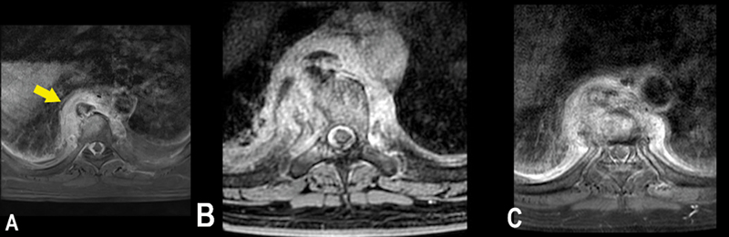
An MRI scan revealed a paravertebral lesion with enhancement of D6 to D9, involving the D7 and D8 vertebrae ([Figs. 2A], [B], [C], and [3A]). However, it was unclear whether the findings were indicative of spondylodiscitis or of a secondary implant. Bone scintigraphy demonstrated localized increase in osteoblastic activity from D6 to D9, with no other sites of increased uptake ([Fig. 4]).


Biological material for the pathological analysis was collected through video-assisted thoracoscopy. The anatomopathological examination revealed inflammatory cells without evidence of neoplasia or infectious agents ([Fig. 5]). Peripheral blood cultures obtained during the patient's admission resulted in the identification of methicillin-resistant Staphylococcus aureus (MRSA), and the diagnosis of spondylodiscitis was established.

The patient remained hospitalized for 6 weeks postdiagnosis, continuing treatment with broad-spectrum antimicrobial drugs and showing clinical improvement. However, subsequent imaging studies showed progression of the paravertebral lesion with involvement of the vertebrae and the intervertebral disc at D7 to D8 ([Figs. 2A], [B], [C] and [3A], [B]). He was discharged with oral antimicrobial sulfamethoxazole-trimethoprim for 60 days, chosen according to the blood culture antibiogram.
One month postdischarge, follow-up imaging studies showed partial resolution of the that the paravertebral lesion. Nevertheless, chronic signs of spondylodiscitis persisted, such as subchrondral irregularities at D7 and D8 and a slight reduction in their heights ([Figs. 2] and [3]). The patient has resumed his activities without any limitations, and he is currently being monitored, with no evidence of disease recurrence.
Discussion
The diagnosis of pain in oncology patients can be challenging, as immunosuppression and multiple approaches during diagnosis and treatment can increase the risk of many pathologies, such as inflammation and infections. Therefore, a thorough clinical evaluation is essential before attributing pain to recurrence or progressive neoplastic disease.
Spondylodiscitis, an infectious condition, often requires prolonged hospitalization and complete rest for the patient. If not promptly recognized and treated, it can progress to chronic pain and irreversible neurological deficits.[5] [10]
The patient in the case herein reported aligns with the epidemiological profile of spondylodiscitis, considering gender, age, DM2, obesity, and neoplasia.[3] [10] The pain may manifest as mechanical or inflammatory in nature.[8] [9] [20] The most frequent pain location is the lumbar region (59%), followed by the thoracic (34%), cervical (10%), and sacral (3%) regions.[1] [6] [10] [21] The laboratory findings were in consisted with those reported in the literature, including leukocytosis with neutrophilia, elevated CRP, and normochromic and normocytic anemia.[19] [20] In chronic cases, leukocyte levels may be increased in 13 to 60% of the cases.[1] [11]
The radiological alterations in the case herein reported were atypical, complicating the initial diagnosis. Chest CT scans revealed a paravertebral lesion with contrast enhancement but without vertebral or intervertebral disc involvement. Thoracic spine MRI showed a paravertebral and vertebral lesion, hypointense on T1-weighted sequences and enhanced after contrast, without intervertebral disc involvement. This ruled out the possibility of discitis and suggested neoplasia or inflammation/infection. The absence of discitis may occur in the early stage of spondylodiscitis, particularly in immunocompromised patients, as in the present case.[5] [8]
The second MRI scan revealed enhancement in vertebral plateaus and discs, raising the possibility of spondylodiscitis. The lesion's progression was likely secondary to manipulation during video-assisted thoracoscopy. The third MRI scan showed the natural evolution of spondylodiscitis.[20]
Spondylodiscitis can develop endogenously via hematogenous spread or exogenously through contiguity from invasive procedures. Hematogenous spread is the most common route, often associated with sepsis and urinary tract, respiratory or gastrointestinal infections.[1] [6] [8] [20] In the case herein reported, a primary infectious focus was not identified, which occurs in approximately 62.5% of the cases.[5] [8] However, MRSA was identified as the causative organism, suggesting that the potential infection route involved breaches in the skin barrier due to microlesions from radiodermatitis, immunosuppression, and an expansive neoplastic lesion of the anal canal and the adjacent skin. Consequently, hematogenous dissemination through the Batson's vertebral venous system led to bacterial colonization of the vertebrae and paravertebral tissues.12 The most common pathogen for spondylodiscitis is MRSA, identified in 20 to 84% of the cases. Nevertheless, Gram-negative bacteria may be implicated in patients who are immunosuppressed, present DM2, and in intravenous drug users.[3] [7] [10] [19] [21] Blood samples are only positive in 35% of the cases, while the biopsy is diagnostic in 53% of the cases.[5]
In the case herein reported, antibiotics were selected considering the patient's risk factors, such as DM2, neoplasia, and recent and prolonged hospitalization. Linezolid is particularly indicated for the treatment of MRSA infections.[11] Clindamycin, when administered in various combinations for an average duration of 30.3 days, has demonstrated efficacy in more than 90% of the cases of spondylodiscitis without an identified pathogen. Carbapenems offer superior Gram-negative coverage compared to other beta-lactams, and they are stable against extended-spectrum beta-lactamases and AmpC beta-lactamases.[2]
The combination of linezolid and clindamycin targeted Gram-positive bacteria, including MRSA. Meropenem broadened the antibiotic range to include Gram-negative bacteria. The patient's ongoing antibiotic treatment may have influenced the negative culture results from the biopsy. The treatment should include intravenous broad-spectrum antibiotics for 6 to 8 weeks until pathogen identification, followed by oral antibiotics for about 6 weeks until laboratory and radiological improvement.[1] [3] [5] [7] [8] The optimal duration of the antibiotic therapy is debated, but therapy lasting fewer than 4 weeks of duration is associated with a 25% recurrence rate.[1] A reduction in CRP ≥ 50%, along with pain attenuation and absence of neurological deficits, are predictors of a switch to oral therapy.[21] In the case herein reported, meropenem therapy was continued due to the patient's status of DM2, immunosuppression, and recent prolonged hospitalization.
Oral antibiotics such as clindamycin, fluoroquinolones, metronidazole, rifampicin, and linezolid are recommended to treat osteomyelitis as alternatives to intravenous antibiotics.[21] These agents should be prioritized for their excellent bone penetration.[7] They also offer excellent bioavailability, high efficacy, minimal toxicity, and reductions in intravenous access and hospitalization time.[21]
Ratiu et al.[2] suggest vancomycin as a potential first-line empirical therapy, along with piperacillin-tazobactam or meropenem. New-generation cephalosporins have recently been shown to be effective in the treatment of MRSA strains.[2] Aminoglycosides exhibit excellent activity against members of the Enterobacteriaceae family and S. aureus, including MRSA.[2] Trimethoprim-sulfamethoxazole is effective against Gram-positive and -negative bacteria, including MRSA, and it demonstrates adequate bone penetration when used at high doses. It was selected based on the antibiogram results in the case herein reported.
The criteria for hospital discharge in spondylodiscitis cases include the absence of back pain, resolution of the inflammatory pattern, normal body temperature, normalized CRP values and/or erythrocyte sedimentation rate, along with stabilization or improvement of disc and vertebrae abnormalities.[10] Magnetic resonance imaging should be used to assess the treatment response, or if a paravertebral abscess is suspected.[8] [10] In a retrospective cohort analysis, Hecquet et al[22]. found that the frequency of soft-tissue infiltration on follow-up radiological imaging was significantly lower than on diagnostic MRI imaging (p < 0.01).[20] Despite the apparent radiological worsening, the patient herein described was discharged due to clinical improvement. The images were taken at short intervals in order to monitor the patient's atypical case.
In conclusion, the evaluation and investigation of pain in oncological patients should consider diagnoses beyond metastasis, such as those of infectious diseases, which are more common due to immunosuppression, increased vulnerability, and the multiple interventions to which this group is submitted.
Conflict of Interests
The authors have no conflict of interests to declare.
Ethics and Procedures
Informed consent was obtained from the patient whose case is being reported in this manuscript. The present case report was authorized by the institutional Ethics Committee. These documents are available in the [Supplementary Material].
Authors' Contributions
All authors should have made substantial contributions to the manuscript. MBRR: collection and assembly of data, data analysis and interpretation, and manuscript writing; LGSR: collection and assembly of data, and manuscript writing; LBD: collection and assembly of data, conception and design, and manuscript writing; JBS: final approval of the manuscript and provision of study materials or patients; and RCC: data analysis and interpretation, provision of study materials or patients, and final approval of the manuscript.
-
References
- 1 Herrero CFPdS, Nascimento ALd, Cunha RP, Souza JPVd, Nogueira-Barbosa MH, Defino HLA. Infectious spondylodiscitis: Has there been any evolution in the diagnostic and treatment outcomes?. Coluna/Columna 2014; 13 (04) 294-297 10.1590/S1808-18512014130400442
- 2 Ratiu IA, Moisa CF, Ţiburcă L, Hagi-Islai E, Ratiu A, Bako GC. et al Antimicrobial Treatment Challenges in the Management of Infective Spondylodiscitis Associated with Hemodialysis: A Comprehensive Review of Literature and Case Series Analysis. Antibiotics (Basel) 2024; 13 (03) 284
- 3 Álvarez-Narváez AR, Elizalde-Martínez E, Moheno-Gallardo AJ, Lares-Cárdenas LA, López-Valencia J, Torres-González R, Morales-de Los Santos R. Caracterización y asociación clínico bacteriológica en la espondilodiscitis piógena. Acta Ortop Mex 2019; 33 (03) 141-145
- 4 Carvalho VNd, Silva FF, Sarmento PM, Baptista S, Sá J. Espondilodiscite Infeciosa: Formas de Apresentação, Diagnóstico e Tratamento. Med Interna 2018; 25 (02) 85-90 10.24950/rspmi/original/218/2/2018
- 5 Faria R, Borges C, Carrondo H, Banza MJ. [Spondylodiscitis: which etiology?]. Acta Med Port 2011; 24 (06) 1059-1064
- 6 Sánchez PM. Spondylodiscitis. Radiologia 2016; 58 (Suppl. 01) 50-59
- 7 Cordero-Delgado DA, Moheno-Gallardo AJ, Torres-González R, Mata-Hernández A, Elizalde-Martínez E, Pérez-Atanasio JM. [Evidence and recommendation of empirical antimicrobial treatment in pyogenic spondylodiscitis: systematic review]. Rev Med Inst Mex Seguro Soc 2017; 55 (Suppl 1): S6-S13
- 8 Finger G, Cecchini AMdL, Sfreddo E, Cecchini FMdL, Lunardi LW, Nascimento TLd, Falavigna A. Spondylodiscitis Investigation and Therapeutic Protocol: Neurosurgery Service Results. Coluna/Columna 2019; 18 (02) 138-143 10.1590/S1808-185120191802195906
- 9 Capelo J, Carragoso A, Albuquerque C, Mocho ML, Canto-Moreira N. Espondilodiscite Infecciosa: o estudo de quarenta e um casos. Acta Reumatol Port 2007; 32 (03) 255-262
- 10 Soto A, Fica A, Dabanch J, Olivares F, Porte L. Espondilodisicitis Chile. Rev Chilena Infectol 2016; 33 (03) 322-330
- 11 Queiroz JWM, Pereira PCAdA, Figueiredo EG. Espondilodiscite: revisão de literatura. Arq Bras Neurocir 2013; 32 (04) 230-236 10.1055/s-0038-1626020
- 12 Sobottke R, Seifert H, Fätkenheuer G, Schmidt M, Gossmann A, Eysel P. Current diagnosis and treatment of spondylodiscitis. Dtsch Arztebl Int 2008; 105 (10) 181-187
- 13 Rogers JE, Sirisaengtaksin A, Leung M, Morris VK, Xiao L, Huey R. et al Hepatic Metastasectomy in Squamous Cell Carcinoma of the Anal Canal: A Case Series of a Curative Approach. Cancers (Basel) 2023; 15 (15) 3890
- 14 Dewdney A, Rao S. Metastatic squamous cell carcinoma of the anus: time for a shift in the treatment paradigm?. ISRN Oncol 2012; 2012: 756591
- 15 Rao S, Guren MG, Khan K, Brown G, Renehan AG, Steigen SE. et al; ESMO Guidelines Committee. Electronic address: clinicalguidelines@esmo.org. Anal cancer: ESMO Clinical Practice Guidelines for diagnosis, treatment and follow-up*. Ann Oncol 2021; 32 (09) 1087-1100
- 16 Gondal TA, Chaudhary N, Bajwa H, Rauf A, Le D, Ahmed S. Anal Cancer: The Past, Present and Future. Curr Oncol 2023; 30 (03) 3232-3250
- 17 Ajani J, Winter K, Gunderson L, Pedersen J. Benson 3rd AB, Thomas Jr CR, et al. Fluorouracil, mitomycin, and radiotherapy vs. fluorouracil, cisplatin, and radiotherapy for carcinoma of the anal canal: Commentary. JAMA 2008; 299 (16) 1914-1921
- 18 Mitsuura C, Miyamoto Y, Ogawa K, Sawayama H, Toihata T, Harada K. et al Successful Management of Anal Squamous Cell Carcinoma With Liver and Ovary Metastases: A Case Report. In Vivo 2022; 36 (06) 3023-3028
- 19 Cariati P, Ozan DP, Rodriguez SA. Vertebral metastasis from squamous cell carcinoma of the tongue. Front Oral Maxillofac Med 2020; 2 (08) 1-3 10.21037/fomm-20-18
- 20 Crombé A, Fadli D, Clinca R, Reverchon G, Cevolani L, Girolami M. et al. Imaging of Spondylodiscitis: A Comprehensive Updated Review-Multimodality Imaging Findings, Differential Diagnosis, and Specific Microorganisms Detection. Microorganisms 2024; 12 (05) 893
- 21 Haddad N, Ajaz J, Mansour L, Kasemodel R, Jarvis J, Jarad J. et al. A Review of the Clinical Utilization of Oral Antibacterial Therapy in the Treatment of Bone Infections in Adults. Antibiotics (Basel) 2023; 13 (01) 4
- 22 Hecquet S, Verhoeven F, Aubry S, Prati C, Wendlin D, Chirouze C. et al. Interest of follow-up radiological imaging in patients with pyogenic vertebral osteomyelitis. Journal of Clinical Medicine 2021; 10 (12) 2690
Address for correspondence
Publication History
Received: 09 April 2025
Accepted: 28 July 2025
Article published online:
11 December 2025
© 2025. The Author(s). This is an open access article published by Thieme under the terms of the Creative Commons Attribution 4.0 International License, permitting copying and reproduction so long as the original work is given appropriate credit (https://creativecommons.org/licenses/by/4.0/)
Thieme Revinter Publicações Ltda.
Rua Rego Freitas, 175, loja 1, República, São Paulo, SP, CEP 01220-010, Brazil
Myllena Batista Ribeiro Rodrigues, Luís Gustavo Soares Rodrigues, Luísa Barbiero Dutra, Janine Bernardi Soder, Rafael Corrêa Coelho. Case Report: Spondylodiscitis as a Differential Diagnosis of Anal Canal Squamous Cell Carcinoma Bone Metastasis. Brazilian Journal of Oncology 2025; 21: s00451812027.
DOI: 10.1055/s-0045-1812027
-
References
- 1 Herrero CFPdS, Nascimento ALd, Cunha RP, Souza JPVd, Nogueira-Barbosa MH, Defino HLA. Infectious spondylodiscitis: Has there been any evolution in the diagnostic and treatment outcomes?. Coluna/Columna 2014; 13 (04) 294-297 10.1590/S1808-18512014130400442
- 2 Ratiu IA, Moisa CF, Ţiburcă L, Hagi-Islai E, Ratiu A, Bako GC. et al Antimicrobial Treatment Challenges in the Management of Infective Spondylodiscitis Associated with Hemodialysis: A Comprehensive Review of Literature and Case Series Analysis. Antibiotics (Basel) 2024; 13 (03) 284
- 3 Álvarez-Narváez AR, Elizalde-Martínez E, Moheno-Gallardo AJ, Lares-Cárdenas LA, López-Valencia J, Torres-González R, Morales-de Los Santos R. Caracterización y asociación clínico bacteriológica en la espondilodiscitis piógena. Acta Ortop Mex 2019; 33 (03) 141-145
- 4 Carvalho VNd, Silva FF, Sarmento PM, Baptista S, Sá J. Espondilodiscite Infeciosa: Formas de Apresentação, Diagnóstico e Tratamento. Med Interna 2018; 25 (02) 85-90 10.24950/rspmi/original/218/2/2018
- 5 Faria R, Borges C, Carrondo H, Banza MJ. [Spondylodiscitis: which etiology?]. Acta Med Port 2011; 24 (06) 1059-1064
- 6 Sánchez PM. Spondylodiscitis. Radiologia 2016; 58 (Suppl. 01) 50-59
- 7 Cordero-Delgado DA, Moheno-Gallardo AJ, Torres-González R, Mata-Hernández A, Elizalde-Martínez E, Pérez-Atanasio JM. [Evidence and recommendation of empirical antimicrobial treatment in pyogenic spondylodiscitis: systematic review]. Rev Med Inst Mex Seguro Soc 2017; 55 (Suppl 1): S6-S13
- 8 Finger G, Cecchini AMdL, Sfreddo E, Cecchini FMdL, Lunardi LW, Nascimento TLd, Falavigna A. Spondylodiscitis Investigation and Therapeutic Protocol: Neurosurgery Service Results. Coluna/Columna 2019; 18 (02) 138-143 10.1590/S1808-185120191802195906
- 9 Capelo J, Carragoso A, Albuquerque C, Mocho ML, Canto-Moreira N. Espondilodiscite Infecciosa: o estudo de quarenta e um casos. Acta Reumatol Port 2007; 32 (03) 255-262
- 10 Soto A, Fica A, Dabanch J, Olivares F, Porte L. Espondilodisicitis Chile. Rev Chilena Infectol 2016; 33 (03) 322-330
- 11 Queiroz JWM, Pereira PCAdA, Figueiredo EG. Espondilodiscite: revisão de literatura. Arq Bras Neurocir 2013; 32 (04) 230-236 10.1055/s-0038-1626020
- 12 Sobottke R, Seifert H, Fätkenheuer G, Schmidt M, Gossmann A, Eysel P. Current diagnosis and treatment of spondylodiscitis. Dtsch Arztebl Int 2008; 105 (10) 181-187
- 13 Rogers JE, Sirisaengtaksin A, Leung M, Morris VK, Xiao L, Huey R. et al Hepatic Metastasectomy in Squamous Cell Carcinoma of the Anal Canal: A Case Series of a Curative Approach. Cancers (Basel) 2023; 15 (15) 3890
- 14 Dewdney A, Rao S. Metastatic squamous cell carcinoma of the anus: time for a shift in the treatment paradigm?. ISRN Oncol 2012; 2012: 756591
- 15 Rao S, Guren MG, Khan K, Brown G, Renehan AG, Steigen SE. et al; ESMO Guidelines Committee. Electronic address: clinicalguidelines@esmo.org. Anal cancer: ESMO Clinical Practice Guidelines for diagnosis, treatment and follow-up*. Ann Oncol 2021; 32 (09) 1087-1100
- 16 Gondal TA, Chaudhary N, Bajwa H, Rauf A, Le D, Ahmed S. Anal Cancer: The Past, Present and Future. Curr Oncol 2023; 30 (03) 3232-3250
- 17 Ajani J, Winter K, Gunderson L, Pedersen J. Benson 3rd AB, Thomas Jr CR, et al. Fluorouracil, mitomycin, and radiotherapy vs. fluorouracil, cisplatin, and radiotherapy for carcinoma of the anal canal: Commentary. JAMA 2008; 299 (16) 1914-1921
- 18 Mitsuura C, Miyamoto Y, Ogawa K, Sawayama H, Toihata T, Harada K. et al Successful Management of Anal Squamous Cell Carcinoma With Liver and Ovary Metastases: A Case Report. In Vivo 2022; 36 (06) 3023-3028
- 19 Cariati P, Ozan DP, Rodriguez SA. Vertebral metastasis from squamous cell carcinoma of the tongue. Front Oral Maxillofac Med 2020; 2 (08) 1-3 10.21037/fomm-20-18
- 20 Crombé A, Fadli D, Clinca R, Reverchon G, Cevolani L, Girolami M. et al. Imaging of Spondylodiscitis: A Comprehensive Updated Review-Multimodality Imaging Findings, Differential Diagnosis, and Specific Microorganisms Detection. Microorganisms 2024; 12 (05) 893
- 21 Haddad N, Ajaz J, Mansour L, Kasemodel R, Jarvis J, Jarad J. et al. A Review of the Clinical Utilization of Oral Antibacterial Therapy in the Treatment of Bone Infections in Adults. Antibiotics (Basel) 2023; 13 (01) 4
- 22 Hecquet S, Verhoeven F, Aubry S, Prati C, Wendlin D, Chirouze C. et al. Interest of follow-up radiological imaging in patients with pyogenic vertebral osteomyelitis. Journal of Clinical Medicine 2021; 10 (12) 2690