
RSS-Feed abonnieren
DOI: 10.1055/s-0040-1722386
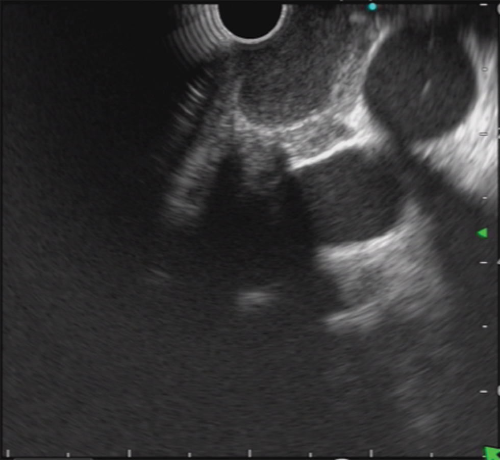
Tubercular Ulcer: Not so Uncommon Cause of Odynophagia


These images are from a 13-year-old girl, who presented with odynophagia. Upper gastrointestinal (GI) endoscopy revealed longitudinal ulcer in the mid esophagus ([Fig. 1]). CT chest [Fig. 2] and endoscopic ultrasound (EUS) ([Fig. 3]) showed a node in the left paratracheal region. Cytology examination showed necrotic granulomas ([Fig. 4]), and stain for acid-fast bacillus (AFB) was positive. Midesophageal ulcers have varied etiology such as viral diseases, pill esophagitis, corrosive injury, submucosal lesions, or malignancy. In endemic places, tuberculosis should be considered as differential diagnosis. Subepithelial bulge with ulcer (summit ulcer) has been explained in tuberculosis.


Publikationsverlauf
Artikel online veröffentlicht:
31. Dezember 2020
© 2020. Society of Gastrointestinal Endoscopy of India. This is an open access article published by Thieme under the terms of the Creative Commons Attribution-NonDerivative-NonCommercial-License, permitting copying and reproduction so long as the original work is given appropriate credit. Contents may not be used for commercial purposes, or adapted, remixed, transformed or built upon. (https://creativecommons.org/licenses/by-nc-nd/4.0/).
Thieme Medical and Scientific Publishers Pvt. Ltd.
A-12, Second Floor, Sector -2, NOIDA -201301, India