Introduction
10.1055/a-1885-5664For a long time, the lungs were considered to be an organ that was sonographically
inaccessible, since total reflection and mirroring of sound waves occurs at the tissue-air
boundary. Since the 1990 s, knowledge about the utility of lung artifacts for clinical
practice has grown rapidly, especially in adult medicine. In the meantime, lung ultrasound
has also found its way into pediatrics and the field of neonatology and, especially
in this patient group, holds great potential that has not yet been fully developed.
As a radiation-free imaging technique, it not only enables a reduction in cumulative
radiation exposure, but also relevantly expands diagnostic possibilities. Nevertheless,
pulmonary sonography is not yet widely used in neonatal units, and further scientific
work is urgently needed to improve the evidence. The following article is intended
to highlight the possibilities and limitations of lung ultrasound and to provide the
basic principles for its useful application in neonatology.
Since no differentiation between classic B-lines and comet tail artifacts is made
in the scientific publications on lung ultrasound (LU) in the neonatal period, presumably
for reasons of simplification, these are also grouped here under the term B-lines.
Learning Goals
-
Detection of sonographic characteristics of transitory tachypnea and respiratory distress
syndrome.
-
Understanding the possibilities but also the limitations of pulmonary sonography in
narrowing down differential diagnoses of respiratory insufficiency.
-
Detection of sonographic abnormalities in pulmonary immaturity and bronchopulmonary
dysplasia.
-
Detection of sonographic features of atelectasis/dystelectasis as well as pneumonia.
-
Understanding the pulmonary sonographic features of pneumothorax.
Transitory Tachypnea and Respiratory Distress Syndrome
Transitory Tachypnea and Respiratory Distress Syndrome
Pathophysiologically, transitory tachypnea (TTN) focuses on increased interstitial
fluid content of the lungs due to delayed reabsorption. Therefore, TTN manifests sonographically
by increased visualizable B-lines. Depending on disease severity, well-separated or
confluent B-lines up to bilateral white lung can be detected, but no relevant consolidations
can be visualized [1]
[2]
[3]
[4]
[5]
[6]
[7]. When first described, evidence of condensed or confluent B-lines in the inferior
fields with few B-lines in the superior fields was considered the most important sonographic
criterion for TTN ([Fig. 1a, ]
[Table 1]). Copetti et al. called the sharp transition a “double-lung point” ([Fig. 1a]) [8]. However, according to current studies, this can be detected in fewer than 50 %
of patients with a clinical diagnosis of TTN, sometimes only during convalescence
[1].
 Fig. 1 Transient tachypnea and respiratory distress syndrome. a Parasternal longitudinal section in a 36th gestational week (GW) preterm infant with transient tachypnea (TTN) after primary
caesarean section (FiO2 0.25 under noninvasive respiratory support). The ultrasound
depicts the usual image of TTN with double lung point (arrowhead). The upper fields
show few B-lines; confluent B-lines are present in the lower fields. The sharp transition
is called the double lung point (arrowhead). b Longitudinal paravertebral section in a 27th week preterm infant with respiratory distress syndrome (FiO2 0.75 under non-invasive
respiratory support). A ubiquitous white lung without visible A-lines is evident.
The subpleural region shows a hypoechoic band with hyperechoic air reflexes corresponding
to consolidated lung tissue.
Fig. 1 Transient tachypnea and respiratory distress syndrome. a Parasternal longitudinal section in a 36th gestational week (GW) preterm infant with transient tachypnea (TTN) after primary
caesarean section (FiO2 0.25 under noninvasive respiratory support). The ultrasound
depicts the usual image of TTN with double lung point (arrowhead). The upper fields
show few B-lines; confluent B-lines are present in the lower fields. The sharp transition
is called the double lung point (arrowhead). b Longitudinal paravertebral section in a 27th week preterm infant with respiratory distress syndrome (FiO2 0.75 under non-invasive
respiratory support). A ubiquitous white lung without visible A-lines is evident.
The subpleural region shows a hypoechoic band with hyperechoic air reflexes corresponding
to consolidated lung tissue.
Table 1
Overview of possible differential diagnoses in respiratory insufficiency according
to sonographic primary findings [1]
[2]
[3]
[4]
[5]
[6]
[7]
[8]
[22]
[23]
[24]
[25]
[26]
[27]
[28]
[31]
[32]
[33]
[34]
[35]
[36]
[37]
[38]
[39].
It should be noted that mixed patterns are common, especially in extremely preterm
infants, but also in more mature preterm infants.
|
Primary finding: increased B-lines
|
|
A-lines/B-lines
|
Pleural line
|
Consolidations
|
Features
|
|
Transitory tachypnea
|
Bilateral B-lines up to white lung
|
Largely unremarkable
|
No relevant consolidations, i. e., expansion < 0.5 cm
|
Possibly double lung point
|
|
Respiratory distress syndrome
|
Bilateral, homogeneously distributed, mostly confluent B-lines
Mostly bilateral white lung
|
Thickened, irregular, altered coarse- or fine-grained
Changes often extend beyond the region of the pleural line
|
Extent dependent on disease severity
|
Main finding dependent on disease severity
Possibly visible lung pulse
|
|
Lung immaturity
|
Increased B-lines as a classic finding in immature lungs
|
Variable
|
Variable
|
Expected findings depending on the degree of lung immaturity (GW, postnatal age) and
current oxygen demand
|
|
Interstitial pulmonary edema e. g., due to hyperhydration, PDA, heart failure, etc.
|
Homogeneously distributed, increased, possibly confluent B-lines.
|
Rather unremarkable
|
Generally none
|
|
|
Primary finding: increased B-lines and consolidation
|
|
A-lines/B-lines
|
Pleural line
|
Consolidations
|
Features
|
|
Respiratory distress syndrome
|
Bilateral, homogeneously distributed, mostly confluent B-lines
Mostly bilateral white lung
|
Thickened, irregular, altered coarse- or fine-grained
Changes often extend beyond the region of the pleural line
|
Extent dependent on disease severity
|
Main finding dependent on disease severity
Possibly visible lung pulse
|
|
Bronchopulmonary dysplasia
|
Increased B-lines
often arising from pleural pathologies or consolidations
|
Irregular, possibly coarsely fragmented
|
Multiple microconsolidations often in combination with more extensive consolidations
(depth > 0.5 cm)
|
As the patient matures, the sonographic abnormalities become more subtle
|
|
Interstitial/atypical pneumonia
|
Increased, diffusely distributed B-lines
|
Irregular, thickened,
fragmented
|
Diffuse microconsolidations
|
|
|
Meconium aspiration syndrome
|
Diffuse, inhomogeneously distributed B-lines
|
Irregular, thickened,
fragmented
|
Diffuse consolidation
|
Adjacent affected and unremarkable areas
|
|
Neonatal ARDS
|
Diffuse, inhomogeneously distributed B-lines
|
Irregular, thickened,
fragmented pleural line
|
Diffuse consolidations increasing in depth and extent with disease severity
|
Adjacent affected and unremarkable areas
|
|
Primary finding: Consolidation
|
|
A-lines/B-lines
|
Pleural line
|
Consolidations
|
Features
|
|
Atelectasis
|
No A-lines in atelectasis region
Possibly B-lines emanating from the edges of the atelectasis
|
Pleura visceralis directly evident
|
Largely anaerobic consolidation
|
Smooth border in atelectasis of a whole lobe of the lung
If extensive atelectasis, midline shift to the side of the atelectasis possible
|
|
Dystelectasis
|
No A-lines in area of pleural dystelectasis
Possibly B-lines emanating from the edges of the dystelectasis
|
Absent or fragmented in the area of the dystelectasis
|
Consolidation with substantial bronchial and possibly alveolar residual air
Irregular deep margins
Dynamic air bronchogram possible
|
Only dystelectases adjacent to the pleura can be visualized
|
|
“Typical” pneumonia / lobar pneumonia
|
No A-lines in the area of pneumonic infiltrates
Consolidation surrounded by increased B-lines
Remaining lung variable
|
Absent or fragmented in the area of dystelectasis
|
Varying residual air distribution
Dynamic air bronchogram typical
|
Mostly irregular deep margins
Possible concomitant effusion
Abscess: inhomogeneous, roundish area with absent perfusion
Only processes adjacent to the pleura can be visualized
|
|
Pulmonary embolism
|
Possibly B-lines emanating from the edges of the consolidation(s)
Remaining lung variable
|
Interrupted in the region of the embolism
|
Generally microconsolidation(s) without residual air
|
Lack of blood flow within the consolidation(s)
|
|
Pulmonary sequestration
|
No A-lines in the area of sequestration
Remaining lung variable
|
Interrupted in the area of sequestration
|
Anaerobic consolidation
|
Margin edge smooth
Internal structure inhomogeneous or cystic
Aortal blood supply
Only visible with contact to pleura
|
|
Congenital cystic adenomatoid malformation (CCAM) of the lung
|
Possibly B-lines emanating from the edges of the consolidation(s)
Wholly variable
|
Variable
|
Consolidation possible as sole feature
Micro-consolidations possible with B-lines emanating from them as sole feature
|
Internal structure inhomogeneous or cystic
Possible reduced/absent pleural sliding
Ultrasound detection of aerated cysts may not be possible
In case of large malformations, midline shift to the opposite side possible
|
|
Primary finding: Absent or diminished pleural sliding
|
|
A-lines/B-lines
|
Pleural line
|
Consolidations
|
Features
|
|
Pneumothorax
|
Clear distinct A-lines in the area of the pneumothorax
|
Smooth, unremarkable in the area of the pneumothorax
|
None in the area of the pneumothorax
|
No pleural sliding
Evidence of lung point in the case of partial pneumothorax
No lung pulse in M-mode
Barcode/stratosphere sign in M-mode
Ventral clear reflection of the rib cartilage
In the case of tension pneumothorax, possible midline shift to the opposite side
|
|
(Massive) Hyperinflation
|
More A-lines than normally expected
|
Variable
|
Consolidations possible
|
Pleural sliding reduced or absent depending on severity
Sonographic detection is currently not possible with certainty
|
|
Misintubation,
tube obstruction, tube dislocation
|
Rapidly reduced or absent A-lines and increasing B-lines
|
Rapid changes to the pleural line
|
Increasing consolidation
In bullous emphysema, the under-ventilation becomes visible with a delay
|
Bilateral absent or significantly reduced pleural sliding
Pleural sliding for determining tube position can only be used reliably in the absence
of spontaneous breathing
|
|
Unilateral endobronchial intubation
|
In non-ventilated areas rapidly reduced or absent A-lines and increasing B-lines
|
In non-ventilated areas rapid changes to the pleural line
|
Rapid formation of consolidations in non-ventilated areas
In bullous emphysema, the under-ventilation becomes visible with a delay
|
Absent pleural sliding in non-ventilated areas
Regular pleural sliding in ventilated areas
Pleural sliding for determining tube position can only be used reliably in the absence
of spontaneous breathing
|
|
Bullae
|
Variable
Possibly well-depicted A-lines in the area of bullae close to the pleura
|
Variable
|
Concomitant microconsolidations possible (especially in cases of concomitant interstitial
emphysema)
|
Reduced/absent pleural sliding in this area is possible in the case of large bullae
close to the pleura
Sonographic detection is currently not possible with certainty
|
Increased B-lines are usually defined in lung ultrasound by the detection of more
than 2 B-lines per intercostal space in the longitudinal scan. In neonatology, this
definition is supplemented by the presence of two or more adjacent intercostal spaces
with confluent B-lines in each lung area examined [6]. Since even lung-healthy premature infants and neonates show increased B-lines during
postnatal adaptation, the detection of increased B-lines in the first days of life
has clinical relevance only in the presence of concomitant pulmonary symptoms [9].
In respiratory distress syndrome (RDS), deficient production of surfactant surface-active
protein leads to inadequate reduction of surface tension with alveolar collapse and
formation of atelectasis. In addition, hypoxia and unphysiologically high opening
pressures lead to lesions of the alveolar epithelia and transfer of a fribinous exudate
into the alveoli with formation of hyaline membranes [10]
[11]. Thus, the pathology is characterized not only by increased interstitial fluid content,
but also by decreased ventilation and inflammatory reactions of the immature lung.
Sonographically, therefore, RDS is characterized by bilateral, homogeneously distributed,
confluent B-lines, a thickened, irregularly coarse or fine-grained altered pleural
line and consolidations ([Fig. 1b], [Video 1], [Table 1]). In higher-grade RDS, a visible lung pulse is also seen in the moving image ([Video 1]) [1]
[4]
[5]
[6]
[7]
[12]. The most important feature of RDS as opposed to TTN is the evidence of consolidation
and the absence of inconspicuous areas ([Fig. 1b, ]
[Video 1], [2a]). As the severity of the RDS increases, the consolidations increase in extent and
depth [2].
Video 1 Respiratory distress syndrome. Longitudinal dorsal section of a quadruplet premature
infant at 27 GW with respiratory distress syndrome (FiO2 0.40 under invasive ventilation).
Sonographically, there is a white lung with a coarse, irregular pleural line. These
changes extend to approximately 0.3–0.4 cm in depth and correspond to poorly aerated
lung tissue. A visible lung pulse is evident in the moving picture.
Video 2 Persistent pulmonary arterial hypertension (PPHN). a Ventral longitudinal section in a preterm infant at 34 + 0 GW at the 5th hour of
life with an oxygen demand of 90 % under noninvasive respiratory support. Clearly
visible A-lines are seen with pleural sliding. In addition, single, predominantly
separated, basally minimally confluent B-lines can be seen, which move synchronously
with pleural sliding. This age-normal pulmonary sonographic finding makes a primary
pulmonary oxygenation disorder with an oxygen demand of 90 % unlikely. Echocardiography
confirmed the suspicion of PPHN. Increasing oxygenation impairment made timely endotracheal
intubation and mechanical ventilation necessary. b Control ultrasound on day 7 of life at 50 % oxygen demand with continued PPHN under
invasive ventilation. Densely packed, homogeneously distributed B-lines are seen with
hardly visible A-lines. In addition, individual microconsolidations limited to the
subpleural region are evident. The worsening of findings is most likely explained
by several days of invasive ventilation in the presence of existing pulmonary immaturity.
Another factor could be persistent ductus arteriosus with cross shunt and pulmonary
flooding.
Differentiation of the two pathologies is not always clear-cut in clinical practice.
Mixed patterns occur especially in more mature preterm infants. Severe, persistent
TTN may also cause secondary surfactant inactivation with alveolar collapse and formation
of consolidations, so that differentiation between TTN and mild RDS is not always
clinically possible with sufficient certainty [2]
[13]
[14]. Several research groups studying sonographic aspects of TTN and RDS developed semiquantitative
lung ultrasound scores (LUS) to objectify the visual impression ([Fig. 2]) [2]
[12]
[15]
[16]
[17]
[18]. Scoring systems allow better comparability of lung ultrasound findings, make interpretation
less dependent on the experience of the examiner, and thus enable integration of the
LU into clinical algorithms. Recent studies demonstrate a close correlation between
ultrasound lung findings, neonatal lung disease severity and oxygenation parameters.
Lung ultrasound scores thus show high predictive value for failure of noninvasive
respiratory support as well as subsequent surfactant application [2]
[12]
[15]
[17]
[18]
[19]
[20].
 Fig. 2 Semiquantitative lung ultrasound score (LUS) according to Brat et al. In the supine
position, each lung region (a total of 6 lung areas with 3 areas per side: anterior
superior, anterior inferior, lateral) is evaluated according to a point system (0–3
points per region). The individual points are summed to a total score. a Score 0: Unremarkable region with well-depicted A-lines. b Score 1: Region with an average of more than 2 well-defined B-lines per intercostal
space. This also applies to inhomogeneous lung regions which have areas with confluent
B-lines as well as inconspicuous sections. The overall impression must be considered
in very small premature infants with a small diameter of the intercostal spaces. c Score 2: Region with confluent B-lines with or without microconsolidations. d Score 3: Region with extensive consolidations with a depth > 0.5 cm.
Fig. 2 Semiquantitative lung ultrasound score (LUS) according to Brat et al. In the supine
position, each lung region (a total of 6 lung areas with 3 areas per side: anterior
superior, anterior inferior, lateral) is evaluated according to a point system (0–3
points per region). The individual points are summed to a total score. a Score 0: Unremarkable region with well-depicted A-lines. b Score 1: Region with an average of more than 2 well-defined B-lines per intercostal
space. This also applies to inhomogeneous lung regions which have areas with confluent
B-lines as well as inconspicuous sections. The overall impression must be considered
in very small premature infants with a small diameter of the intercostal spaces. c Score 2: Region with confluent B-lines with or without microconsolidations. d Score 3: Region with extensive consolidations with a depth > 0.5 cm.
The score used in most studies was published by Brat et al. in 2015 ([Fig. 2]). Implementation of this score in ward guidelines for surfactant therapy has resulted
in significantly earlier surfactant administration with lower inspiratory oxygen delivery
without increasing the rate of application in subsequent intervention studies when
the cut-off value is > 8 [16]
[21]. The score according to Brat et al. has been modified and applied in many studies
by various research groups. The modifications in this case affect both the pulmonary
areas studied and the definitions of the individual point values, which makes it difficult
to calculate cut-off values in meta-analyses. Despite divergent scoring systems, a
correlation in the same direction between the total score and the inspiratory oxygenation
required for adequate ventilation was consistently demonstrated. Thus, as the score
increases, the gas exchange capacity of the lung deteriorates, from which a direct
correlation can be inferred between sonographic findings and the severity of lung
injury. Lung ultrasound scoring systems represent the future of sonographically guided,
individualized therapy regimens, although further standardization and adaptation to
different levels of maturity would be desirable for widespread use.
The correlation between the level of a pulmonary-related oxygen demand and sonographic
findings also helps to differentiate a predominantly pulmonary from an extrapulmonary
etiology in cases of persistently high postnatal oxygenation. If oxygen demand and
sonographic findings do not coincide, other causes, including extrapulmonary causes,
such as persistent pulmonary arterial hypertension ([Video 2a]), vitium cordis, and, of course, pneumothorax ([Video 3]), as well as hyperinflation, must be included in the differential diagnostic considerations.
Video 3 Pneumothorax. Ventral longitudinal section in a preterm infant at 30 + 4 GW with
an oxygen demand of 60 % under noninvasive respiratory support. Clearly visible A-lines
are seen in the moving image with absent visualization of pleural sliding. On closer
inspection, the movements below the tissue-air boundary correspond merely to reflections
of the movements of the thoracic wall structures above the tissue-air boundary. No
B-lines can be seen.
Bronchopulmonary Dysplasia
Bronchopulmonary Dysplasia
Much less is known regarding the further development of pulmonary sonographic pathologies
of preterm infants than the early postnatal period. Only since 2021 has the number
of publications on this topic increased noticeably.
In the further course of the disease, multifactorial influencing variables – such
as the time of sufficient surfactant synthesis, fluid status, patent ductus arteriosus
(PDA) and pulmonary inflammatory reactions due to oxygen exposure, mechanical ventilation
or sepsis – increasingly determine the ultrasound lung findings ([Video 2b]). The rectified relationship between respiratory insufficiency and LUS continues
steadily, so that there is always a correlation between sonographic lung status and
an existing oxygenation disturbance ([Fig. 3], [4], [5]) [22]
[23]. In addition to the correlation with oxygenation parameters independent of gestational
age, lung ultrasound findings always show a dependence on the degree of lung immaturity,
which is determined by gestational age at birth and postnatal age [22]
[23]. Typically, the score deteriorates within the first week of life ([Fig. 3], [4], [Video 2b]) and then improves continuously. In contrast, in preterm infants with developing
bronchopulmonary dysplasia (BPD), pathologic sonographic phenomena persist in parallel
with oxygen demand ([Fig. 5], [6]). Changes in the pleural line with diffuse microconsolidations and resulting vertical
reverberation artifacts and consolidations are clearly apparent ([Fig. 4], [5], [6]). Extensive consolidations are typically localized in the dorsal lung fields ([Fig. 4]), so that these should be included in the sonographic evaluation in the further
course of the disease. With increasing maturity and decreasing oxygen demand, patients
with bronchopulmonary dysplasia also show improvement in lung sonographic findings
([Fig. 5]), but in preterm infants with moderate and severe BPD, the pathologic changes persist
beyond 36 + 0 gestational weeks ([Fig. 6], [Table 1]) [23]
[24]
[25]
[26]
[27]
[28]. Therefore, for developing or manifest BPD, lung ultrasound represents a promising
additional bedside imaging modality for pulmonary monitoring and a potential parameter
for therapy management.
 Fig. 3 Typical lung ultrasound findings in extremely premature infants with oxygenation
impairment in the first days of life. a+b Bilateral parasternal longitudinal section in a triplet premature infant (2nd triplet, birthweight 375 g) of 22 + 6 gestational weeks on day 3 of life (FiO2 0.28–0.30
under invasive ventilation, immediate postnatal surfactant administration). a A predominantly intact pleural line is seen on the right ventral side. Regions with
single, well separated B-lines with still sporadically visible A-lines are shown next
to areas with confluent B-lines without visible A-lines. b Left ventral band-like consolidations confined to the subpleural region are evident,
giving the appearance of an irregularly fragmented pleural line. The irregular, deep
edges of the under-ventilated zones are starting points of vertical, confluent repeat
echoes. Retrocardial microconsolidations are present. The sonographic findings suggest
that the right ventral lung is better ventilated than the left. Additional findings
include a small, irrelevant pericardial effusion. c Parasternal longitudinal section on the right and longitudinal section at the level
of the anterior axillary line on the left in a triplet premature baby (3rd triplet, birthweight 330 g, surfactant administration immediately postnatally) at
22 + 6 GW on day 3 of life (FiO2 0.30–0.32 under invasive ventilation). Confluent
B-lines without displayable A-lines can be seen in both slice planes. The pulmonary
sonographic findings show extreme pulmonary immaturity with mild to moderate oxygenation
impairment.
Fig. 3 Typical lung ultrasound findings in extremely premature infants with oxygenation
impairment in the first days of life. a+b Bilateral parasternal longitudinal section in a triplet premature infant (2nd triplet, birthweight 375 g) of 22 + 6 gestational weeks on day 3 of life (FiO2 0.28–0.30
under invasive ventilation, immediate postnatal surfactant administration). a A predominantly intact pleural line is seen on the right ventral side. Regions with
single, well separated B-lines with still sporadically visible A-lines are shown next
to areas with confluent B-lines without visible A-lines. b Left ventral band-like consolidations confined to the subpleural region are evident,
giving the appearance of an irregularly fragmented pleural line. The irregular, deep
edges of the under-ventilated zones are starting points of vertical, confluent repeat
echoes. Retrocardial microconsolidations are present. The sonographic findings suggest
that the right ventral lung is better ventilated than the left. Additional findings
include a small, irrelevant pericardial effusion. c Parasternal longitudinal section on the right and longitudinal section at the level
of the anterior axillary line on the left in a triplet premature baby (3rd triplet, birthweight 330 g, surfactant administration immediately postnatally) at
22 + 6 GW on day 3 of life (FiO2 0.30–0.32 under invasive ventilation). Confluent
B-lines without displayable A-lines can be seen in both slice planes. The pulmonary
sonographic findings show extreme pulmonary immaturity with mild to moderate oxygenation
impairment.
 Fig. 4 Worsening of the lung ultrasound findings with increasing oxygen demand. Follow-up
(same patient as in [Fig. 3a, b]) on day 8 of life for respiratory deterioration in the context of focal intestinal
perforation (FiO2 0.60 under invasive ventilation). Ventrally right shows single micro
consolidations as well as predominantly confluent B-lines with barely visualizable
A-lines (a). Ventrally left shows the picture of a sonographically white lung with band-like
decreased ventilation of the subpleural region and retrocardiac microconsolidations
(b). The main finding, however, is bilateral dorsolateral with new onset, deeper consolidations
with positive air bronchogram (c).
Fig. 4 Worsening of the lung ultrasound findings with increasing oxygen demand. Follow-up
(same patient as in [Fig. 3a, b]) on day 8 of life for respiratory deterioration in the context of focal intestinal
perforation (FiO2 0.60 under invasive ventilation). Ventrally right shows single micro
consolidations as well as predominantly confluent B-lines with barely visualizable
A-lines (a). Ventrally left shows the picture of a sonographically white lung with band-like
decreased ventilation of the subpleural region and retrocardiac microconsolidations
(b). The main finding, however, is bilateral dorsolateral with new onset, deeper consolidations
with positive air bronchogram (c).
 Fig. 5 Typical sonographic findings in the further course of hospitalization in extremely
premature infants with persistent oxygenation impairment. a+b Ultrasound examination of a premature infant at 23 + 3 GW (birthweight 410 g) at
8 weeks of age with respiratory global insufficiency (FiO2 0.95–1.0 under invasive
ventilation). Ubiquitous extensive consolidations (atelectasis/dystelectasis) and
increased B-lines are present. Shown here is a ventral cross-section in the area of
the right upper lobe (a) and a right-sided, paravertebral longitudinal section (b). The consolidations reach into the deep regions of the lungs (depth > 1 cm), include
several intercostal spaces and only show some hyperechoic residual air in the deep
areas. The deep edges are irregular and are the origin of vertical artifacts. c+d Check-up after 12 days with an improved respiratory situation (FiO2 0.35 with CPAP
breathing support). Greater reduced ventilation zones are no longer visible. However,
ubiquitous small microconsolidations limited to the subpleural region (reduced ventilation)
are seen, which give rise to the appearance of a grossly fragmented, irregular pleural
line, especially ventrally. The reduced ventilation areas are the starting points
for predominantly well-separated B-lines. Regular A-lines can be seen in regions with
only a few vertical artifacts. This finding persisted in severe BPD in parallel with
oxygen demand beyond 36 + 0 GW.
Fig. 5 Typical sonographic findings in the further course of hospitalization in extremely
premature infants with persistent oxygenation impairment. a+b Ultrasound examination of a premature infant at 23 + 3 GW (birthweight 410 g) at
8 weeks of age with respiratory global insufficiency (FiO2 0.95–1.0 under invasive
ventilation). Ubiquitous extensive consolidations (atelectasis/dystelectasis) and
increased B-lines are present. Shown here is a ventral cross-section in the area of
the right upper lobe (a) and a right-sided, paravertebral longitudinal section (b). The consolidations reach into the deep regions of the lungs (depth > 1 cm), include
several intercostal spaces and only show some hyperechoic residual air in the deep
areas. The deep edges are irregular and are the origin of vertical artifacts. c+d Check-up after 12 days with an improved respiratory situation (FiO2 0.35 with CPAP
breathing support). Greater reduced ventilation zones are no longer visible. However,
ubiquitous small microconsolidations limited to the subpleural region (reduced ventilation)
are seen, which give rise to the appearance of a grossly fragmented, irregular pleural
line, especially ventrally. The reduced ventilation areas are the starting points
for predominantly well-separated B-lines. Regular A-lines can be seen in regions with
only a few vertical artifacts. This finding persisted in severe BPD in parallel with
oxygen demand beyond 36 + 0 GW.
 Fig. 6 Severe bronchopulmonary dysplasia. Ultrasound examination of the lungs in a twin
premature baby of 24 + 6 GW (birthweight 700 g) with severe bronchopulmonary dysplasia
at 36 + 0 GW (FiO2 0.42 under high-flow). a+b Ventrally, there are multiple, small, underventilated zones near the pleura, which
form the image of a coarsely fragmented, irregular pleural line. Vertical artifacts
emanate from these poorly aerated zones; A-lines are visible in open regions. c+d Dorsally, there are under-ventilated areas on both sides, some of which are > 0.5 cm
deep and extend over several intercostal spaces. Vertical artifacts emanate from the
under-ventilated zones; A-lines are visible in open areas.
Fig. 6 Severe bronchopulmonary dysplasia. Ultrasound examination of the lungs in a twin
premature baby of 24 + 6 GW (birthweight 700 g) with severe bronchopulmonary dysplasia
at 36 + 0 GW (FiO2 0.42 under high-flow). a+b Ventrally, there are multiple, small, underventilated zones near the pleura, which
form the image of a coarsely fragmented, irregular pleural line. Vertical artifacts
emanate from these poorly aerated zones; A-lines are visible in open regions. c+d Dorsally, there are under-ventilated areas on both sides, some of which are > 0.5 cm
deep and extend over several intercostal spaces. Vertical artifacts emanate from the
under-ventilated zones; A-lines are visible in open areas.
Some studies also demonstrate a predictive value of sonographic scoring systems for
the development of moderate or severe BPD as early as between the seventh and fourteenth
days of life [23]
[24]
[25]
[26]
[27]
[28]. Future studies should consider whether pulmonary sonography is superior to clinical
scoring systems in this regard as well as research the significance of pulmonary sonographic
pathologies for long-term pulmonary prognosis.
Atelectasis and Dystelectasis
Atelectasis and Dystelectasis
Lung consolidations in the context of developing or manifest BPD are partly atelectasis
(nonventilated lung areas) and partly dystelectasis (inadequately ventilated lung
areas) ([Fig. 3b], [4], [5], [6], [7]). These can also be detected within the framework of other diseases and especially
in invasively ventilated patients with respiratory failure. Here, sonographically
even the smallest microatelectasis near the pleura due to insufficient respiration
or insufficient ventilation volume can be detected with high sensitivity and the contained
residual air distribution can be accurately assessed ([Fig. 3b], [4], [Video 4], [Table 1]) [29]. Using lung ultrasound, a positioning treatment can therefore be better controlled
than by means of conventional AP X-ray. In addition, ultrasound can be used to monitor
reduced ventilations serially, without radiation ([Fig. 7], [Video 4]).
 Fig. 7 Atelectasis. Dorsal atelectasis in a twin premature infant of 23 + 4 GW (birthweight
490 g) at 4 weeks of age with respiratory global failure (FiO2 0.85 under invasive
HFO ventilation). In the supine position (after repositioning), there are extensive
bilateral dorsal reduced ventilation zones surrounded by confluent B-lines. Longitudinal
section on the right shows longitudinal extension of consolidation from lung apex
to lung base at a depth > 1 cm (a). In cross-section, the main finding is directly paravertebral (b) Only a few, hyperechogenic air reflections can be seen on both planes. During the
course of the day, a reduction of the oxygen supply up to a minimum of 40 % was possible
by supine position and recruitment maneuvers. The preterm infant was able to be discharged
home later with only mild BPD and no need for supplemental oxygen.
Fig. 7 Atelectasis. Dorsal atelectasis in a twin premature infant of 23 + 4 GW (birthweight
490 g) at 4 weeks of age with respiratory global failure (FiO2 0.85 under invasive
HFO ventilation). In the supine position (after repositioning), there are extensive
bilateral dorsal reduced ventilation zones surrounded by confluent B-lines. Longitudinal
section on the right shows longitudinal extension of consolidation from lung apex
to lung base at a depth > 1 cm (a). In cross-section, the main finding is directly paravertebral (b) Only a few, hyperechogenic air reflections can be seen on both planes. During the
course of the day, a reduction of the oxygen supply up to a minimum of 40 % was possible
by supine position and recruitment maneuvers. The preterm infant was able to be discharged
home later with only mild BPD and no need for supplemental oxygen.
Video 4 Lung ventilation improvement through recruitment maneuver. Continuation of examination
in [Fig. 6]. a Clip after prone position for 10–15 min (FiO2 0.80 under invasive HFO ventilation).
As an indication of a free airway and an incipient improvement in ventilation, ultrasound
already shows slightly more residual air in the upper field. b After a 2-minute increase in MAP of 2 mmHg, increasing re-aeration of the atelectatic
areas is seen sonographically. In the middle sections, a pleural line with B-lines
emanating from it and recognizable A-lines can be visualized in 3 intercostal spaces.
Ultrasound alone cannot assess whether local hyperinflation has already occurred in
these areas. In the sections that are still poorly ventilated, more air reflexes can
be seen within the consolidations. Synchronously to the sonographic improvement, the
oxygen demand was reduced from 80 % to 60 % and in the further course of the day to
40 %.
Therefore, in adult medicine, ultrasound is also used for ventilation optimization
and PEEP control [30]. In our own experience, improvements in sonographic findings are also seen in neonatology
after ventilation optimization with decreasing oxygen demand ([Fig. 7], [Video 4]). If ultrasound is used as an aid for ventilation optimization, however, the user
should be aware that while pleural underinflation can be detected and controlled very
sensitively, pulmonary hyperinflation cannot be reliably detected sonographically.
In addition, not all under-ventilation responds to recruitment maneuvers. Since neonatal
patients with chronic lung injury typically have inhomogeneous lungs with a juxtaposition
of overinflated and underinflated areas, this must always be taken into account when
using ultrasound for PEEP or ventilation control to avoid additional exacerbation
of local overinflation.
Pneumonia
Pneumonia with inflammatory infiltrate also presents sonographically as consolidation
surrounded by increased B-lines ([Fig. 8], [Table 1]). In healthy lung patients with increased signs of inflammation, typical clinical
symptoms and sonographically detectable extensive consolidation, the suspected diagnosis
of pneumonia can be confirmed by ultrasound alone ([Fig. 8]) [31]. Sonographic criteria for differentiation between pneumonia and atelectasis are
not applicable with sufficient specificity in neonatology ([Fig. 8], [Video 5]). For example, some authors consider lack of residual air in the bronchial system
as a marker of atelectasis and a dynamic air bronchogram as a criterion for pneumonic
infiltrate ([Fig. 8], [Video 5]). However, inadequate ventilation in preterm infants requiring oxygen is typically
characterized by more or less pronounced residual bronchial and alveolar air, and
dystelectatic consolidations predominate over atelectasis ([Video 5b]). As [Video 5b] demonstrates, a dynamic air bronchogram can occasionally be documented in dystelactatic
regions even in the absence of an inflammatory genesis. In neonatal pneumonia, moreover,
concomitant secretory obstruction occurs rapidly, so that a dynamic air bronchogram
is not necessarily observed here ([Fig. 9]). Thus, pneumonia cannot be differentiated with sufficient certainty from dystelectasis
typical of preterm infants with oxygen demand using sonography alone. In this case,
correct interpretation is only possible in conjunction with the clinical symptoms
and the progression of the disease. However, pneumonia can be very well monitored
sonographically during its course and possible complications such as purulent effusions
or abscess formation can be detected at an early stage [32]
[33].
 Fig. 8 Pneumonia. 3-month-old former 27th GW preterm infant with dyspnea, new-onset oxygen demand and elevated inflammatory
parameters. a Dorsal longitudinal section shows inhomogeneously distributed B-lines on the left,
originating from the margins of a small consolidation (inflammatory reduced ventilation)
(arrow). The rest of the lung shows a regular pleural line and well-depicted A-lines.
b Longitudinal section reveals a large, hypoechoic reduced ventilation zone (pneumonic
infiltrate) in the right dorsal upper lobe with tree-like residual hyperechogenic
air in the bronchial system (arrows). Together with the clinic signs, the diagnosis
of pneumonia can be made.
Fig. 8 Pneumonia. 3-month-old former 27th GW preterm infant with dyspnea, new-onset oxygen demand and elevated inflammatory
parameters. a Dorsal longitudinal section shows inhomogeneously distributed B-lines on the left,
originating from the margins of a small consolidation (inflammatory reduced ventilation)
(arrow). The rest of the lung shows a regular pleural line and well-depicted A-lines.
b Longitudinal section reveals a large, hypoechoic reduced ventilation zone (pneumonic
infiltrate) in the right dorsal upper lobe with tree-like residual hyperechogenic
air in the bronchial system (arrows). Together with the clinic signs, the diagnosis
of pneumonia can be made.
Video 5 Dynamic air bronchogram showing pneumonia and dystelectasis. a Continuation of examination in [Fig. 7]. On closer inspection, respiratory synchronous movements of the air reflexes within
the consolidation (pneumonic infiltrate) can be visualized in the video. b Premature infant at 22 + 6 GW with respiratory deterioration in the setting of intestinal
perforation during strangulation ileus (FiO2 0.60 under invasive ventilation). Lateral
cross-section shows dorsal hypoechoic reduced ventilation (dystelectasis) with dynamic
air bronchogram. Respiratory synchronous movements of the air reflexes can be seen
in different areas of the small airways.
 Fig. 9 Pneumonia. Premature 24 + 1 GW infant with respiratory deterioration on day 35 of
life requiring secondary intubation, viscous tracheal secretions, and CRP elevation
to 6 mg/dl (FiO2 0.80 under invasive ventilation). In dorsal longitudinal as well
as cross-sectional view, there is extensive lung consolidation with little residual
intrabronchial air. Lung tissue shows a liver-like echotexture (hepatization) with
inhomogeneous echogenicity. Taken together with the clinical signs, the findings were
considered pneumonia.
Fig. 9 Pneumonia. Premature 24 + 1 GW infant with respiratory deterioration on day 35 of
life requiring secondary intubation, viscous tracheal secretions, and CRP elevation
to 6 mg/dl (FiO2 0.80 under invasive ventilation). In dorsal longitudinal as well
as cross-sectional view, there is extensive lung consolidation with little residual
intrabronchial air. Lung tissue shows a liver-like echotexture (hepatization) with
inhomogeneous echogenicity. Taken together with the clinical signs, the findings were
considered pneumonia.
Pneumothorax
Pneumothorax (PTX) is always an important differential diagnosis in respiratory failure
during the premature and neonatal period, so it must always be ruled out as a cause,
especially in acute respiratory deterioration. Lung ultrasound also enables rapid
diagnosis directly at the patientʼs bedside in emergencies and has been shown in studies
to be superior to standard radiological diagnostics in sensitivity and specificity
due to better detection of small amounts of air (ventral pneumothorax) [34]
[35]. Thus, ultrasound allows the detection of minimal intrapleural air volumes and should
replace chest X-ray as the gold standard of PTX diagnosis in neonatology due to its
various advantages.
PTX is the result of air entering the pleural space. If PTX leads to total collapse
of the lung, the two pleural sheets no longer touch. In partial PTX, air collects
at the highest point of the pleural cavity depending on the position. Depending on
the extent of the pneumothorax, the visceral pleura and parietal pleura are still
contiguous in more or less large areas ([Video 6], [7]). Free air below the parietal pleura sonographically also results in the visualization
of a delicate hyperechogenic line at the tissue-air interface with multiple horizontal
reverberation artifacts ([Fig. 10], [Video 3], [6], [7]). In contrast, other ultrasound artifacts typical for the lung cannot be detected
([Fig. 10], [Video 3], [6], [7]). Thus, PTX is sonographically characterized by lack of pleural sliding, lack of
visualization of B-lines and consolidations, visualization of the so-called lung point
where the visceral pleura and parietal pleura separate, and lack of visualization
of the pulmonary pulse in M-mode ([Fig. 10], [11]
[Video 1], [6]–[8], [Table 1]) [6]
[34].
Video 6 Pneumothorax. Dorsal longitudinal section in a preterm infant at 27 + 1 GW (FiO2
0.30 under binasal CPAP). No regular pleural sliding can be visualized in the region
of the two cranial intercostal spaces. In the lowest intercostal space, however, a
regular atemsynchronous displacement of the pleural line against the structures of
the thoracic wall is detectable adjacent to the liver. In this area, close inspection
of the tissue-air interface can identify the lung point and diagnose partial pneumothorax.
Video 7 Incremental diagnostics to assess the extent of a pneumothorax. a Longitudinal section at the level of the anterior axillary line on the left in an
extremely premature baby with an oxygen requirement of 50 % on the 2nd day of life and sonographic suspicion of pneumothorax (same patient as in [Fig. 3c, d]). Neither regular pleural sliding nor B-lines or consolidations can be detected.
b Longitudinal section at the posterior axillary line on the left shows a poorly aerated
lung with band-like decreased aeration confined to the subpleural region, confluent
vertical reverberation artifacts emanating from it, and a visible lung pulse. c In lateral cross-section, the lung point can be visualized between the mid and posterior
axillary lines. Thus, the diagnosis is a partial pneumothorax with separation of the
two pleural sheets in the area between the middle and posterior axillary line. As
the oxygen demand continued to increase, prompt placement of a drain was required.
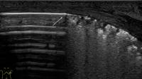 Fig. 10 Left pneumothorax. Pneumothorax in a premature infant 24 GW (FiO2 0.35 under invasive
ventilation). Left lateral intercostal cross-section. Ventral presentation of multiple
reverberation artifacts, corresponding to pneumothorax. On the other hand, there are
two-dimensional microconsolidations and B-lines emanating laterodorsally from them.
The lung point (arrow) is located at the level of the midaxillary line.
Fig. 10 Left pneumothorax. Pneumothorax in a premature infant 24 GW (FiO2 0.35 under invasive
ventilation). Left lateral intercostal cross-section. Ventral presentation of multiple
reverberation artifacts, corresponding to pneumothorax. On the other hand, there are
two-dimensional microconsolidations and B-lines emanating laterodorsally from them.
The lung point (arrow) is located at the level of the midaxillary line.
 Fig. 11 M-mode pneumothorax. Comparison of the seashore sign in regular pleural sliding (a) and the barcode sign in pneumothorax (b) in M-mode. a Regular pleural sliding causes blurred or coarse-grained visualization of dorsally
located repeat echoes. In addition, the lung pulse (asterisk) is formed synchronously
by vibration of the pleural line (arrowhead) and thus the dorsal echoes. b In the absence of pleural sliding in pneumothorax, straight lines appear both above
and below the tissue-air interface (arrowhead). No lung pulse can be detected.
Fig. 11 M-mode pneumothorax. Comparison of the seashore sign in regular pleural sliding (a) and the barcode sign in pneumothorax (b) in M-mode. a Regular pleural sliding causes blurred or coarse-grained visualization of dorsally
located repeat echoes. In addition, the lung pulse (asterisk) is formed synchronously
by vibration of the pleural line (arrowhead) and thus the dorsal echoes. b In the absence of pleural sliding in pneumothorax, straight lines appear both above
and below the tissue-air interface (arrowhead). No lung pulse can be detected.
Video 8 Pneumothorax and transient tachypnea. a Longitudinal parasternal section in a preterm infant at 35 GW with dyspnea and mild
oxygen demand on CPAP respiratory support. In the upper fields, few single B-lines
are seen; in the lower fields, confluent B-lines with small reduced ventilation zones
and visible pulmonary pulse are seen. Regular pleural sliding can be visualized in
all areas. The abrupt transition is the double lung point typical of TTN. b Longitudinal parasternal section in a preterm infant at 31 GW with incompletely drained
pneumothorax. At first glance, the sonographic findings resemble the double-lung point
of TTN shown in a. However, close inspection reveals no pleural sliding cranial to this point. The
movements below the hyperechogenic tissue-air boundary correspond to reflections of
the movements of the thoracic wall structures. Thus, it is the lung point in partial
pneumothorax.
In premature infant and neonate requiring oxygen, PTX is a sonographic visual diagnosis.
Missing B-lines with simultaneously well-visualized A-lines that mainly make the examiner
consider PTX when the transducer is first placed on the highest point of the thorax
([Video 1], [7a]). Nevertheless, the most important sonographic criterion of PTX remains the absence
of pleural sliding, otherwise misdiagnosis may occur. Therefore, an accurate assessment
of the pleural line in the moving image using a high-resolution linear transducer
and a shallow penetration depth should always be performed ([Video 6]). In M-mode, the lack of pleural sliding in pneumothorax does not show the typical
seashore sign but rather the pathognomic barcode or stratosphere sign ([Fig. 11]) [6]
[34].
Assessment of pleural sliding can be complicated by the patientʼs severe respiratory
effort in the presence of dyspnea ([Video 8b]). M-mode does not offer any advantages here, as the motion artifacts limit the differentiation
between seashore sign and barcode sign and may make reliable detection of the missing
lung pulse impossible. If there is no bilateral PTX, comparing the sides in the median
cross-section can help to reliably identify the unilateral absence of pleural sliding.
If the lung point can be found in addition to the missing pleural sliding, the PTX
is considered proven ([Fig. 10], [Video 6], [7]) [6]. At the lung point, the intrapleural air causes the two pleural layers to separate.
On the side of the PTX there is no sliding of the pleura, on the other side of the
lung point regular sliding of the pleura can usually be seen in combination with B-lines
or consolidations ([Fig. 6], [7]). In case of uncertainty, the positional dependence of the intrapleural air and
thus the lung point can be used for confirmation.
Sonographically, no statement can be made about the distance between the visceral
pleura and the parietal pleura. However, the lung point can be used to detect the
boundaries of the PTX and assess its extent ([Video 7a–c]) [36], thus allowing repeated follow-ups without radiation. In the supine position, the
reference of the lung point to the midaxillary line appears to allow differentiation
between a small “ventral” pneumothorax and pneumothorax requiring treatment. Ultimately,
however, the decision to place a drain should always be based on the clinical condition
of the patient.
If neither B-lines, subpleural pathology, nor regular pleural sliding can be demonstrated
in well-depicted A-lines, and if no pulmonary point can be demonstrated, the diagnosis
is most likely total collapse of the lung in pneumothorax. In case of doubt – and
time permitting – this diagnosis can be confirmed by the absence of a lung pulse in
M-mode ([Fig. 9b]) [6]. In addition, sometimes in the supine position, the completely collapsed lung can
be visualized far dorsally.
The most important sonographic differential diagnosis to PTX in the neonatal period
represents the image of a TTN with double lung point ([Video 8]). It is imperative to definitively differentiate the lung point of the PTX from
the double lung point of the TTN ([Video 8]). The most important distinguishing feature is the pleural sliding, which can be
detected regularly in all sides in TTN ([Video 8]). Differential diagnosis must also take into account extrapulmonary causes of high
oxygen demand with unremarkable pulmonary imaging. It should always be kept in mind
that after pleurodesis or in the presence of pleural adhesions of other causes, the
assessment of pleural sliding can no longer be safely used for PTX diagnosis. Also,
if cutaneous emphysema is present, assessment of the dorsally located structures is
no longer possible. In cutaneous emphysema or mediastinal pneumothorax, the artifact-causing
gas is located in the plane of the skin or mediastinum, respectively, so that precise
localization allows differentiation from intrapleural or intrapulmonary air.
Further Differential Diagnoses
Further Differential Diagnoses
In the case of acute respiratory failure, lung ultrasound can also help in the clinical
context with other pathologies in a “rule in-rule out” procedure to quickly narrow
down possible differential diagnoses at the bedside. [Table 1] provides overview of the most important neonatal differential diagnoses [1]
[2]
[3]
[4]
[5]
[6]
[7]
[8]
[22]
[23]
[24]
[25]
[26]
[27]
[28]
[31]
[32]
[33]
[34]
[35]
[36]
[37]
[38]
[39]. However, it must be taken into account that mixed pictures are often present especially
in very immature preterm infants with chronic lung pathology.
Conclusions
In neonatology, lung ultrasound is an excellent imaging modality for bedside serial
assessment of pulmonary pathology, delineation of differential diagnoses, and detection
of complications. The close relationship between clinical and sonographic pulmonary
status suggests direct visualization of underlying pathological changes using ultrasound,
thus making lung ultrasound an ideal monitoring tool in the clinical framework. When
integrated into the ward routine as a point-of-care procedure, not only can a significant
reduction in cumulative radiation exposure be achieved, but also an improvement in
treatment quality. In differential diagnostic considerations, clinical symptoms, laboratory
findings and sonographic image must always be considered in concert. The most complete
possible observation of the lung surface improves diagnostic certainty, especially
in the later course of the disease. In neonatology, however, relevant lung pathologies,
which currently cannot be detected with sufficient certainty by ultrasound, also play
a role. These include, in particular, pulmonary hyperinflation and pulmonary interstitial
as well as bullous emphysema. The user should always be aware of these limitations
and consult other imaging techniques to clarify these issues. Perhaps in the future,
innovative sonographic techniques such as special perfusion imaging, contrast-enhanced
ultrasound (CEUS), tissue Doppler and elastography can fill the existing gaps and
further improve the diagnostic confidence of lung ultrasound.


