

Subscribe to RSS
DOI: 10.1055/s-0035-1552619
Infective Causes of Epilepsy
Authors
Abstract
A wide range of infections of the central nervous system are responsible for both acute seizures and epilepsy. The pathogenesis and clinical semiology of the seizure disorders vary widely between the infective pathogens. The exact mechanisms underlying this are poorly understood, but appear, at least in part, to relate to the pathogen; the degree of cortical involvement; delays in treatment; and the host inflammatory response. The treatment of infective causes of seizures involves both symptomatic treatment with antiepileptic drugs and direct treatment of the underlying condition. In many cases, early treatment of the infection may affect the prognosis of the epilepsy syndrome. The greatest burden of acute and long-term infection-related seizures occurs in resource-poor settings, where both clinical and research facilities are often lacking to manage such patients adequately. Nevertheless, education programs may go a long way toward addressing the stigma, leading to improved diagnosis, management, and ultimately to better quality of life.
Infectious Agents Causing Epilepsy
Any infection of the cortex can potentially result in seizures. This primarily relies upon the structural damage occurring during the infection, although secondary inflammatory processes may also provoke seizures. Certain conditions, including viral encephalitis and parasitic infections—such as “cerebral” malaria and neurocysticercosis—are inherently associated with seizures and the risk of developing epilepsy. Other conditions, such as bacterial meningitis, can cause seizures in the acute setting, although this is less frequent and they rarely result in long-term epilepsy. Here we will review the most common central nervous system (CNS) infectious disorders causing epilepsy, specifically focusing on viral encephalitis and parasitic infections.
Viral Encephalitis
Encephalitis encompasses a broad range of pathophysiological process that result in inflammation of brain parenchyma and therefore the diagnosis is fundamentally a histopathological one.[1] [2] In view of the impracticality of obtaining brain tissue, surrogate markers of brain inflammation are routinely used to make the diagnosis. These include elevated cerebrospinal fluid (CSF) leukocyte counts, raised protein, evidence of viral nucleic acid in the CSF by polymerase chain reaction (PCR), or intrathecal synthesis of antiviral antibody. Neuroimaging can also help ([Fig. 1]).[1] [2] Encephalitis usually presents with an encephalopathy syndrome for which there is often a broad differential diagnosis.[3] Features raising suspicion for infective encephalitis include a history of fever or coryzal illness, recent travel, and inadequate vaccination.[1] A wide range of pathogens can cause encephalitis. Antibody-associated encephalitis, which may be paraneoplastic or de novo, is of increasingly recognized importance, but it is beyond the scope of this article and is covered elsewhere in this themed edition.

Encephalitis results in roughly 19,000 hospitalizations and 1,400 deaths per year in the United States[4] with a reported incidence between 0.7 to 13.8 per 100,000 per year.[1] [3] [5] It is an important condition as delays in diagnosis and in starting treatment can result in significant morbidity and mortality. Acute symptomatic seizures occur in 2 to 67% of patients with a diagnosis of encephalitis overall.[4] The incidence of acute symptomatic seizures and the subsequent development of epilepsy vary with the cause of encephalitis, the patient's comorbidities, delays in starting treatment, and the degree of cortical inflammation.[6] Epidemiological studies of seizures and the long-term outcome for encephalitis are limited and available data are mainly from passive surveillance; therefore, they are prone to underestimate, particularly in resource-poor settings.
Patients with encephalitis risk developing seizures in the acute setting, but are also more likely to develop later unprovoked seizures as described by the International League Against Epilepsy (ILAE).[7] Overall, patients with encephalitis are around 16 times more likely than the general population to develop epilepsy.[4] Of those who have seizures during the acute encephalitic illness, the risk is higher at a 22 times more likely. Even those who do not suffer from acute symptomatic seizures during the acute viral encephalitis are 10 times more likely to develop later unprovoked seizures. The annual incidence of epilepsy due to encephalitis in the United States is approximately the same as that due to head injuries, even though there are 1.4 million cases of CNS trauma per year, compared with fewer than 50,000 cases of CNS infection.[4]
Most commonly, seizures develop within the first 5 years of the acute encephalitic illness, but they may occur up to 20 years after the acute event. The pathogen may be important in determining whether a person subsequently develops epilepsy in the long term. For example, encephalitis caused by La Crosse virus has a cumulative incidence of epilepsy of 10 to 12% while Nipah virus encephalitis has an incidence of approximately 2.2%.[4]
Causes of Infectious Encephalitis
Acute infections causing encephalitis group broadly into those occurring either sporadically or epidemically. The most common sporadic cause is herpes simplex virus, usually type 1, although 10% are type 2.[1] Others include varicella zoster virus and enteroviruses. Epidemic encephalitis most commonly occurs in geographically restricted seasonal epidemics and is caused by arthropod-borne viruses (arboviruses), although clinicians need to be vigilant to the potential for advances in the geographical distribution of these pathogens. Globally, the most important epidemic cause is Japanese encephalitis virus, while others include West Nile virus, tick-borne encephalitis virus and Nipah virus ([Table 1]).[5]
|
Groups |
Viruses |
Comments |
|---|---|---|
|
Sporadic causes |
||
|
Herpes viruses (Family Herpesviridae) |
Herpes simplex virus type 1 |
Most commonly diagnosed sporadic encephalitis |
|
Herpes simplex virus type 2 |
Causes meningitis in adults; meningoencephalitis typically occurs in the immunocompromised; radiculitis |
|
|
Varicella zoster virus |
Postinfective cerebellitis, or acute infective encephalitis or vasculopathy |
|
|
Epstein–Barr virus |
Encephalitis in the immunocompromised |
|
|
Cytomegalovirus |
Encephalitis in the immunocompromised; retinitis, radiculitis |
|
|
Human herpes viruses 6 and 7 |
Febrile convulsions in children; encephalitis in the immunocompromised |
|
|
Enteroviruses (Family Picornaviridae) |
Enterovirus 70 |
Epidemic hemorrhagic conjunctivitis, with CNS involvement |
|
Enterovirus 71 |
Epidemic hand, foot, and mouth disease, aseptic meningitis, brainstem encephalitis, myelitis |
|
|
Poliovirus |
Myelitis |
|
|
Coxsackie viruses, echoviruses, parechoviruses |
Aseptic meningitis |
|
|
Paramyxoviruses (Family Paramyxoviridae) |
Measles virus |
Acute postinfective encephalitis, subacute encephalitis, subacute sclerosing panencephalitis |
|
Mumps virus |
Parotitis, orchitis, pancreatitis are associated with meningoencephalitis |
|
|
Others (rarer) |
Influenza viruses, adenovirus, parvovirus B19, lymphocytic choriomeningitis virus, rubella virus |
|
|
Arthropod-borne and zoonotic viruses [a] |
||
|
Flaviviruses (Family Flaviviridae) |
West Nile virus |
North America, Southern Europe, Middle East, West and Central Asia associated with flaccid paralysis and parkinsonian movement disorders |
|
Japanese encephalitis virus |
Asia, associated with flaccid paralysis and parkinsonian movement disorders |
|
|
Tick-borne encephalitis virus |
Eastern Europe, Former USSR; upper limb flaccid paralysis |
|
|
Dengue viruses (types 1–4) |
Causes fever, arthralgia, rash and hemorrhagic disease; occasional CNS disease |
|
|
Alphaviruses (Family Togaviridae) |
Western, Eastern and Venezuelan equine encephalitis viruses |
Found in the Americas; associated with human and equine encephalitis |
|
Chikungunya virus |
Asia Pacific, Africa |
|
|
Bunya viruses |
La Crosse Virus |
Encephalitis in America |
|
Coltiviruses |
Colorado tick fever virus |
North America |
|
Rhabdoviruses |
Rabies, virus other lyssaviruses |
Non-arthropod-borne zoonotic viruses transmitted by dogs, cats, bats depending on location |
|
Chandipura virus |
Transmitted by sand-flies, causing outbreaks in India |
|
|
Henipaviruses |
Nipah virus |
Transmitted in feces of fruit bats in Malaysia, Bangladesh |
Source: Adapted with permissions from Michael BD, Solomon T. Seizures and encephalitis: clinical features, management, and potential pathophysiologic mechanisms. Epilepsia 2012;53(Suppl 4):63–71.
Abbreviations: CNS, central nervous system.
a Most are zoonotic with the exception of dengue and chikungunya viruses. Viral causes of chronic encephalitis like JC virus are not included.
Herpes Simplex Virus Encephalitis
Herpes simplex virus (HSV) encephalitis has an annual incidence of 1 in 250,000 to 500,000, although this may be an underestimate.[8] Herpes simplex virus type 1 is an α herpes double-stranded DNA virus with which most people are infected by adulthood.[8] It is transmitted by droplet spread and crosses the mucous membranes of the naso-oral cavity and then travels by retrograde axonal transport to sensory ganglia, predominantly the trigeminal ganglion. There it achieves latency, but reactivates periodically, traveling by anterograde axonal transport to the cutaneous nerve endings, resulting in spread of the virus, in some cases associated with a localized vesicular eruption, “herpes labialis.” Triggers for viral reactivation include stress, local trauma, fever, immunosuppression, menstruation, or hormone imbalance. The mechanisms resulting in latency and reactivation are not fully understood, but toll-like receptors and the type 1 and 2 interferon response probably play a crucial role in initially establishing and subsequently maintaining latency.[9] Rarely, the virus replicates in the brain, causing encephalitis although it is unclear whether this result from further retrograde axonal transport after reactivation in the trigeminal ganglion, or whether this is due to reactivation of HSV already latent within the brain ([Fig. 2]).

Herpes simplex virus type 1 encephalitis has a bimodal age distribution, with one-third of cases occurring in young adults and one-half of cases occurring in people older than 50 years.[10] Seizures occur in approximately 40% of people with HSV 1 encephalitis during the acute illness and may be the presenting symptom.[11] The seizures are typically focal, with and without cognitive involvement, reflecting temporal lobe and to a lesser extent frontal lobe infection. Clinical features that typically precede seizures include alterations in cognition, consciousness, personality, or behavior, in the context of an intercurrent or recent febrile illness.[3] However, approximately 11% of patients with HSV encephalitis are afebrile on admission.[12] Associated clinical features include headache, nausea, vomiting, and features suggesting meningism and raised intracranial pressure.[12] The presence of acute symptomatic seizures was associated with a worse outcome in a series of patients with HSV encephalitis; furthermore, the development of status epilepticus is associated with a worse outcome in encephalitis more broadly.[4] [13] In addition, a greater impairment in consciousness, older age, and a delay in starting acyclovir treatment of > 48 hours are each associated with a worse outcome.[4]
Approximately 45 to 60% of people with HSV encephalitis develop epilepsy later in life.[4] [5] [8] Initial treatment should be with an antiepileptic medication effective for focal-onset seizures, although these patients frequently require multiple antiepileptic medications.[5] If a patient has early recurrent seizures, evolving seizure types—particularly with a faciobrachial dystonic semiology—or has associated progressive cognitive decline or neuropsychiatric symptoms, clinicians should consider an early relapse of the encephalitis. This may be due to resurgent viral infection or a postinfective primarily immunomediated process that may be associated with specific antibodies directed against CNS antigens.[13] Early relapses may develop in up to 10 to 26% of patients, especially those who received insufficient acyclovir.[11] However, patients occasionally develop relapses up to 2 years after the initial event.[14] Therefore, UK guidelines recommend that immunocompetent patients older than 12 years should receive intravenous acyclovir for 14 days, and then undergo a repeat HSV PCR of the CSF.[1] [2] If there is still evidence of HSV infection, they should receive a another 7 days of treatment with further CSF sampling thereafter, and acyclovir continued until the CSF PCR is negative. These guidelines also recommend that a minimum treatment duration of 21 days for immunocompromised patients or those younger than 12 years before repeating CSF sampling.[1] [2]
Japanese Encephalitis Virus[15]
An estimated 70,000 cases of encephalitis due to Japanese encephalitis virus (JEV) occur per year, causing 20,500 deaths.[16] Although responsible for seasonal epidemics mostly centered around South East Asia, some cases also occur in the Western Pacific and Eastern Mediterranean.[17] Japanese encephalitis virus is a flavivirus that has a zoonotic transmission with infection between a wide range of vertebrates by Culex mosquitoes, and the predominant cycle is between wading birds, such as egrets, and pigs ([Fig. 3]). Pigs act as amplifying hosts due to high, prolonged viremia.[16] This cycle can spill over to humans who are a dead-end host following a mosquito bite. Therefore, the major risk factors for JEV infection are close proximity to rice fields and pigsties.[18] Acute infection results in a transient, nonspecific febrile viremic illness; in some people, the virus crosses the blood–brain barrier resulting in viral replication in the CNS. This leads to leucocyte transfer into the brain with perivascular cuffing.[17] Treatment is generally supportive and includes management of the complications such as seizures and raised intracranial pressure. Importantly, Japanese encephalitis is a vaccine-preventable illness; thus, delivering broad and comprehensive vaccination programs is of utmost priority.[19]

Japanese encephalitis virus causes acute symptomatic seizures in up to 85% of children and in 10% of adults.[20] [21] Generalized tonic–clonic seizures are more frequent than focal motor seizures; however, the clinical signs may be subtle and seizures can be easily missed, especially if there is limited access to electroencephalography (EEG). In one large cohort study, half of the patients with generalized tonic–clonic seizures and all of those with subtle seizures were in status epilepticus.[21] Moreover, of patients with confirmed encephalitis due to JEV, those who had acute symptomatic seizures while hospitalized were at least 4 times more likely to have a poor outcome.[21] Around half of patients who survive the acute illness are left with severe neurologic sequelae, including motor impairment, and cognitive and language dysfunction.[22] [23] Approximately 20% of survivors are thought to develop long-term seizures, but we need more detailed studies assessing longer-term consequences with a particular focus on late unprovoked seizures and the health economic and social implications.[5]
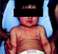
Role of Human Herpes Virus 6
The relatively recently identified lymphotropic β herpes DNA virus human herpes virus 6 (HHV6) is now realized to be the most common cause of roseola infantum in children aged under 2 years ([Fig. 4]).[24] More broadly, HHV6 may account for 20% of emergency department admissions for all febrile illnesses in children aged 6 to 12 months, of whom approximately13% may develop acute seizures.[25] In one study of children aged below 5 years presenting with febrile status epilepticus to an emergency department, 32% had HHV-6B viremia.[26] HHV6 is found by PCR of the CSF in 10% of cases of suspected viral CNS disease, regardless of age.[27] In addition, four studies have reported HHV6 viral DNA in some samples of resected hippocampi from people with mesial temporal lobe sclerosis. HHV6 viral DNA was found in nine (28%) hippocampal samples that were resected for mesial temporal lobe epilepsy in a Chinese study, but only in one (8%) of the control patients.[28] In other studies, there was HHV6 DNA in 15 (62.5%), 4 (50%), and 3 (9%) patients with mesial temporal sclerosis, although there was no control group.[29] [30] [31] This wide variability may reflect patient populations sampled; clearly, we need further work on a larger well-defined cohort with an appropriate control group. In addition, as HHV6 viral DNA can show chromosomal integration, the finding of viral DNA in CSF or brain parenchyma needs confirmation of its significance, for example, through establishing a high or rising viral load, an intrathecal antibody response, and excluding chromosomal integration.

Parasitic Infections
Seizures can complicate parasitic infections of the CNS and may develop both as an acute phase response as well as long-term consequence, although establishing the causality of some organisms can be difficult ([Table 2]).[32] Seizures develop through several mechanisms, including local pressure effects from parasitic growth in the brain parenchyma (e.g., hydatid disease), from inflammation after larval migration, or as part of a diffuse encephalopathy (e.g., African trypanosomiasis). Two major infections associated with a high frequency of seizures and epilepsy are cerebral malaria and neurocysticercosis.[30]
|
Type of lesion |
Agent |
|---|---|
|
Focal granulomatous/cystic lesions |
Protozoa: Toxoplasma, free living amoeba, Chagas disease Nematodes: Toxocariasis, gnathostoma Trematodes: Schistosomiasis, paragonimiasis Cestodes: Cysticercosis, hydatid disease, coenurosis, sparganosis |
|
Abscess |
Entamoeba histolytica, toxoplasmosis |
|
Encephalitis |
Chagas disease, free-living amoebas, “cerebral malaria” |
|
Meningitis |
Angiostrongylus, cysticercosis |
|
Mass effect |
Cysticercosis, hydatid disease, coenurosis |
Cerebral Malaria
There are an estimated 1 million deaths attributed to malaria per year—with half of these resulting from cerebral malaria—of whom 90% are children.[33] The World Health Organization has defined cerebral malaria as unexplained coma in patients with a parasitemia without any other explanation.[34] In addition, cerebral malaria may be suspected clinically by a malaria-specific retinopathy, which is 95% sensitive and 90% specific for children who are comatose secondary to cerebral malaria.[35] The epidemiology and clinical phenotype of the condition varies by age and geographical location. Cerebral disease most commonly develops with falciparum malaria and affects mostly African children below the age of 5 years with a rapid onset of coma and focal motor seizures. Adults normally develop cerebral dysfunction in conjunction with dysfunction of other organs.
Up to 30% of African children have some neurologic sequelae following cerebral malaria; up to 10% develop epilepsy.[36] [37] The mechanism by which cerebral malaria results in epilepsy is not fully understood, but it probably involves a parasite-driven inflammatory process causing cortical damage, which may predominantly affect the occipital neocortex[38] and can be confirmed as focal atrophy on neuroimaging months to years later.[39] However, one case report described hippocampal sclerosis and mesial temporal lobe epilepsy following cerebral malaria.[40] Clinical seizures are occasionally generalized at onset, but more often are focal with or without subsequent secondary generalization; approximately 20% of children have subclinical seizures.[36] Moreover, status epilepticus can occur in up to 28% of patients and having acute seizures is associated with an increased risk of both mortality and long-term neurologic disability.[33] [36] [41]
Treatment involves standard antimalarial medication and best supportive care, which may require intensive care input where available. Therefore, clinicians should be vigilant for the potential for seizures in patients with cerebral malaria, and should consider an EEG, where available, to detect subtle motor or nonconvulsive status epilepticus, especially in those with altered and/or fluctuating consciousness. However, even having identified status epilepticus, the optimal treatment regimen is unclear. In one study, neither phenytoin nor fosphenytoin controlled status epilepticus in 36 to 64% of children with cerebral malaria, despite therapeutic antiepileptic blood concentrations.[41]
Neurocysticercosis
Neurocysticercosis is the infection of the CNS by the larval stage of the tapeworm Taenia solium, and is the most common helminthic infection of CNS globally.[42] Taenia solium is endemic in most developing countries and as such is a major public health problem. Millions of people living in the developing world are infected by the larval form of T. solium, and many of these will experience some degree of clinical consequence of the infection.[43] It is rare in northern Europe, Canada, Australia, Japan, New Zealand, and in many countries with a predominant Muslim population. However, immigration from endemic countries has increased the incidence in these countries more recently. The increasingly widespread use of cysticidal drugs, improved sanitation, and public health educational measures have been associated with a decreasing prevalence of clinical infection. However, neurocysticercosis remains an important cause of hospital admission and a major cause for acquired epilepsy in many areas.[44] [45]
The life cycle of T. solium involves humans, as the only definite hosts for the adult “cestode,” and pigs, which act in conjunction with humans as intermediate hosts for the larval form or cysticercus. The parasite's lifecycle passes through four stages including the vesicular stage, the colloidal stage, the granular stage, and finally the calcified stage ([Fig. 5]).

Approximately 80% of patients with symptomatic neurocysticercosis develop recurrent seizures. Other manifestations include focal neurologic deficits, increased intracranial pressure, and cognitive decline.[46] Neurocysticercosis is a leading cause of acquired epilepsy in endemic areas and is partly responsible for the increased prevalence of epilepsy in the developing world. These patients probably have focal-onset seizures with rapid generalization. Seizures occur predominantly when the parasites begin to degenerate; calcified lesions have been considered to be inert. However, recent reports suggest that calcified lesion may cause seizures in some people when parasitic antigens trapped in the calcium matrix are exposed to the host immune system during calcification remodeling ([Fig. 6]).[47] Neurocysticercosis has also been associated with hippocampal sclerosis.[48] In an appropriate patient with acute symptomatic seizures and exposure to an endemic region, T. Solium can be detected on a simple plain radiograph of the quadriceps ([Fig. 7]).


Management of Seizures
At the moment, no evidence supports the use of antiepileptic medication as primary prophylaxis in patients with a CNS infection. Indeed, there is limited evidence to guide treatment of seizures that occur during or after the acute CNS infection.[1] [2] [6] [49] There have been no randomized or quasirandomized controlled trials for the primary or secondary prophylaxis of seizures in viral encephalitis; therefore, there is currently no evidence to support primary prophylaxis, although secondary prevention is routine practice.[49] A further complicating factor is the management of iatrogenic respiratory failure in the setting of limited resources, as identified in one study of phenobarbital in patients with cerebral malaria.[50] We need large multicenter randomized controlled trials, but before this, we need risk stratification tools to identify those at high risk of developing seizures and status epilepticus.
Convulsive status epilepticus should be treated in the same way as that due noninfective causes, but depends upon local resources. It is important to keep in mind parainfectious processes that complicate brain infections, including cerebral venous sinus thrombosis and electrolyte abnormalities such as hyponatremia. Nonconvulsive status epilepticus should be excluded in comatose patients, particularly if there is fluctuation in consciousness and/or subtle motor features.
Epilepsy following CNS infections remains a challenge especially in resource-poor settings. The treatment, to some extent, depends upon the natural history of the disease;, at the moment, there are no clear predictors of postinfective epilepsy.[4] [11] In HSV encephalitis the timing of antiviral medication can reduce the short-term mortality; however, there are few data assessing early antiepileptic medication and the prevention of long-term sequelae.[51] [52] Regardless of the cause, seizure treatment is targeted toward seizure semiology, although many patients with seizures following CNS infections require a combination of antiepileptic medications.
The WHO has produced guidelines specifically targeted at resource-poor settings.[53] These include the assertion that the diagnosis of epilepsy is clinical and that there should not be an overreliance on neuroimaging and EEG. They also state that nonspecialist health care workers should be trained to recognize and to treat convulsive seizures. Patients should be offered monotherapy with any standard antiepileptic drug—which can be made available sustainably—and the WHO recommends phenobarbital for patients with generalized-onset and carbamazepine for focal-onset epilepsy. Other first-line medications to consider include lamotrigine, given its efficacy for both focal and generalized seizures,[54] and levetiracetam, given the low rates of drug interactions and utility in managing convulsive status epilepticus (provided sustainable availability can be achieved).[55]
Charities, such as the Encephalitis Society, can provide support for patients and their family globally, including supplying information.[1] In resource-poor settings, education programs directed at the patient, their family, and the wider community as to the nature of epilepsy and the basic management of seizures are pivotal in addressing the social stigma that can significantly limit adherence and seizure control, result in injury, and more broadly impact on quality of life.[53]
Conclusion
Infections of the CNS are an important cause of both acute seizures and later epilepsy. The exact mechanisms underlying this are poorly understood. However, they appear, at least in part, to relate to the pathogen, the degree of cortical involvement, delays in treatment, and the host inflammatory response. The greatest burden of acute and long-term infection-related seizures occurs in resource-poor settings, where there may be inadequate clinical and research facilities to manage such patients. Nevertheless, education programs may go a long way toward addressing the damaging stigma, and so lead to improved diagnosis, management, and ultimately quality of life.
Acknowledgments
BDM received Doctoral Fellowship funding from the National Institute for Health Research (NIHR) and is a NIHR Academic Clinical Lecturer. TS received support from the NIHR Health Protection Research Unit in Emerging and Zoonotic Infections at Liverpool. BDM is a Trustee for the Encephalitis Society and TS is Chair of their Scientific Advisory Panel.
-
References
- 1 Solomon T, Michael BD, Smith PE , et al; National Encephalitis Guidelines Development and Stakeholder Groups. Management of suspected viral encephalitis in adults—Association of British Neurologists and British Infection Association National Guidelines. J Infect 2012; 64 (4) 347-373
- 2 Kneen R, Michael BD, Menson E , et al; National Encephalitis Guidelines Development and Stakeholder Groups. Management of suspected viral encephalitis in children - Association of British Neurologists and British Paediatric Allergy, Immunology and Infection Group national guidelines. J Infect 2012; 64 (5) 449-477
- 3 Michael BD, Sidhu M, Stoeter D , et al; North West Neurological Infections Network. Acute central nervous system infections in adults—a retrospective cohort study in the NHS North West region. QJM 2010; 103 (10) 749-758
- 4 Misra UK, Tan CT, Kalita J. Viral encephalitis and epilepsy. Epilepsia 2008; 49 (Suppl. 06) 13-18
- 5 Michael BD, Solomon T. Seizures and encephalitis: clinical features, management, and potential pathophysiologic mechanisms. Epilepsia 2012; 53 (Suppl. 04) 63-71
- 6 Misra UK, Kalita J. Seizures in encephalitis: predictors and outcome. Seizure 2009; 18 (8) 583-587
- 7 Berg AT, Berkovic SF, Brodie MJ , et al. Revised terminology and concepts for organization of seizures and epilepsies: report of the ILAE Commission on Classification and Terminology, 2005-2009. Epilepsia 2010; 51 (4) 676-685
- 8 Whitley RJ. Herpes simplex encephalitis: adolescents and adults. Antiviral Res 2006; 71 (2-3) 141-148
- 9 Widener RW, Whitley RJ. Herpes simplex virus. In: Alex CT, John B, , eds. Handbook of Clinical Neurology. Vol 123. Amsterdam, The Netherlands: Elsevier; 2014: 251-263
- 10 Tyler KL. Herpes simplex virus infections of the central nervous system: encephalitis and meningitis, including Mollaret's. Herpes 2004; 11 (Suppl. 02) 57A-64A
- 11 Solomon T, Hart IJ, Beeching NJ. Viral encephalitis: a clinician's guide. Pract Neurol 2007; 7 (5) 288-305
- 12 Granerod J, Ambrose HE, Davies NW , et al; UK Health Protection Agency (HPA) Aetiology of Encephalitis Study Group. Causes of encephalitis and differences in their clinical presentations in England: a multicentre, population-based prospective study. Lancet Infect Dis 2010; 10 (12) 835-844
- 13 Höftberger R, Armangue T, Leypoldt F, Graus F, Dalmau J. Clinical neuropathology practice guide 4-2013: post-herpes simplex encephalitis: N-methyl-D aspartate receptor antibodies are part of the problem. Clin Neuropathol 2013; 32 (4) 251-254
- 14 De Tiège X, Rozenberg F, Des Portes V , et al. Herpes simplex encephalitis relapses in children: differentiation of two neurologic entities. Neurology 2003; 61 (2) 241-243
- 15 de Bilbao F, Arsenijevic D, Moll T , et al. In vivo over-expression of interleukin-10 increases resistance to focal brain ischemia in mice. J Neurochem 2009; 110 (1) 12-22
- 16 Campbell GL, Hills SL, Fischer M , et al. Estimated global incidence of Japanese encephalitis: a systematic review. Bull World Health Organ 2011; 89 (10) 766-774 , 774A–774E
- 17 Griffiths MJ, Turtle L, Solomon T. Japanese encephalitis virus infection. Handb Clin Neurol 2014; 123: 561-576
- 18 Impoinvil DE, Solomon T, Schluter WW , et al. The spatial heterogeneity between Japanese encephalitis incidence distribution and environmental variables in Nepal. PLoS ONE 2011; 6 (7) e22192
- 19 Solomon T. New vaccines for Japanese encephalitis. Lancet Neurol 2008; 7 (2) 116-118
- 20 Kumar R, Mathur A, Kumar A, Sharma S, Chakraborty S, Chaturvedi UC. Clinical features & prognostic indicators of Japanese encephalitis in children in Lucknow (India). Indian J Med Res 1990; 91: 321-327
- 21 Solomon T, Dung NM, Kneen R , et al. Seizures and raised intracranial pressure in Vietnamese patients with Japanese encephalitis. Brain 2002; 125 (Pt 5) 1084-1093
- 22 Kumar R, Mathur A, Singh KB , et al. Clinical sequelae of Japanese encephalitis in children. Indian J Med Res 1993; 97: 9-13
- 23 Huy BV, Tu HC, Luan TV, Lindqvist R. Early mental and neurological sequelae after Japanese B encephalitis. Southeast Asian J Trop Med Public Health 1994; 25 (3) 549-553
- 24 Campadelli-Fiume G, Mirandola P, Menotti L. Human herpesvirus 6: An emerging pathogen. Emerg Infect Dis 1999; 5 (3) 353-366
- 25 Hall CB, Long CE, Schnabel KC , et al. Human herpesvirus-6 infection in children. A prospective study of complications and reactivation. N Engl J Med 1994; 331 (7) 432-438
- 26 Epstein LG, Shinnar S, Hesdorffer DC , et al; FEBSTAT study team. Human herpesvirus 6 and 7 in febrile status epilepticus: the FEBSTAT study. Epilepsia 2012; 53 (9) 1481-1488
- 27 Studahl M, Hagberg L, Rekabdar E, Bergström T. Herpesvirus DNA detection in cerebral spinal fluid: differences in clinical presentation between alpha-, beta-, and gamma-herpesviruses. Scand J Infect Dis 2000; 32 (3) 237-248
- 28 Li J-M, Lei D, Peng F , et al. Detection of human herpes virus 6B in patients with mesial temporal lobe epilepsy in West China and the possible association with elevated NF-κB expression. Epilepsy Res 2011; 94 (1-2) 1-9
- 29 Fotheringham J, Donati D, Akhyani N , et al. Association of human herpesvirus-6B with mesial temporal lobe epilepsy. PLoS Med 2007; 4 (5) e180
- 30 Donati D, Akhyani N, Fogdell-Hahn A , et al. Detection of human herpesvirus-6 in mesial temporal lobe epilepsy surgical brain resections. Neurology 2003; 61 (10) 1405-1411
- 31 Karatas H, Gurer G, Pinar A , et al. Investigation of HSV-1, HSV-2, CMV, HHV-6 and HHV-8 DNA by real-time PCR in surgical resection materials of epilepsy patients with mesial temporal lobe sclerosis. J Neurol Sci 2008; 264 (1-2) 151-156
- 32 Kamuyu G, Bottomley C, Mageto J , et al; Study of Epidemiology of Epilepsy in Demographic Sites (SEEDS) group. Exposure to multiple parasites is associated with the prevalence of active convulsive epilepsy in sub-Saharan Africa. PLoS Negl Trop Dis 2014; 8 (5) e2908
- 33 Postels DG, Birbeck GL. Cerebral malaria. Handb Clin Neurol 2013; 114: 91-102
- 34 Severe falciparum malaria. World Health organization, communicable diseases cluster. Trans R Soc Trop Med Hyg 2000; 94 (Suppl. 01) S1-S90
- 35 Lewallen S, Wills BA. Retinal haemorrhage in children with malaria. Lancet 1993; 341 (8842) 442
- 36 Birbeck GL, Molyneux ME, Kaplan PW , et al. Blantyre Malaria Project Epilepsy Study (BMPES) of neurological outcomes in retinopathy-positive paediatric cerebral malaria survivors: a prospective cohort study. Lancet Neurol 2010; 9 (12) 1173-1181
- 37 Ngoungou EB, Poudiougou B, Dulac O , et al. [Persistent neurological sequelae due to cerebral malaria in a cohort of children from Mali]. Rev Neurol (Paris) 2007; 163 (5) 583-588
- 38 Opoka RO, Bangirana P, Boivin MJ, John CC, Byarugaba J. Seizure activity and neurological sequelae in Ugandan children who have survived an episode of cerebral malaria. Afr Health Sci 2009; 9 (2) 75-81
- 39 Potchen MJ, Birbeck GL, Demarco JK , et al. Neuroimaging findings in children with retinopathy-confirmed cerebral malaria. Eur J Radiol 2010; 74 (1) 262-268
- 40 Schijns OE, Visser-Vandewalle V, Lemmens EM, Janssen A, Hoogland G. Surgery for temporal lobe epilepsy after cerebral malaria. Seizure 2008; 17 (8) 731-734
- 41 Ogutu BR, Newton CR, Muchohi SN , et al. Pharmacokinetics and clinical effects of phenytoin and fosphenytoin in children with severe malaria and status epilepticus. Br J Clin Pharmacol 2003; 56 (1) 112-119
- 42 Del Brutto OH. Neurocysticercosis. Handb Clin Neurol 2014; 121: 1445-1459
- 43 Ndimubanzi PC, Carabin H, Budke CM , et al. A systematic review of the frequency of neurocyticercosis with a focus on people with epilepsy. PLoS Negl Trop Dis 2010; 4 (11) e870
- 44 Medina MT, Aguilar-Estrada RL, Alvarez A , et al. Reduction in rate of epilepsy from neurocysticercosis by community interventions: the Salamá, Honduras study. Epilepsia 2011; 52 (6) 1177-1185
- 45 Sotelo J, Diaz-Olavarrieta C. Neurocysticercosis: changes after 25 years of medical therapy. Arch Med Res 2010; 41 (1) 62-63
- 46 Carabin H, Ndimubanzi PC, Budke CM , et al. Clinical manifestations associated with neurocysticercosis: a systematic review. PLoS Negl Trop Dis 2011; 5 (5) e1152
- 47 Nash TE, Pretell EJ, Lescano AG , et al; Cysticercosis Working Group in Peru. Perilesional brain oedema and seizure activity in patients with calcified neurocysticercosis: a prospective cohort and nested case-control study. Lancet Neurol 2008; 7 (12) 1099-1105
- 48 Bianchin MM, Velasco TR, Takayanagui OM, Sakamoto AC. Neurocysticercosis, mesial temporal lobe epilepsy, and hippocampal sclerosis: an association largely ignored. Lancet Neurol 2006; 5 (1) 20-21
- 49 Pandey S, Rathore C, Michael BD. Antiepileptic drugs for the primary and secondary prevention of seizures in viral encephalitis. Cochrane Database Syst Rev 2014; 10: CD010247
- 50 Crawley J, Waruiru C, Mithwani S , et al. Effect of phenobarbital on seizure frequency and mortality in childhood cerebral malaria: a randomised, controlled intervention study. Lancet 2000; 355 (9205) 701-706
- 51 Sköldenberg B, Forsgren M, Alestig K , et al. Acyclovir versus vidarabine in herpes simplex encephalitis. Randomised multicentre study in consecutive Swedish patients. Lancet 1984; 2 (8405) 707-711
- 52 Whitley RJ, Alford CA, Hirsch MS , et al. Vidarabine versus acyclovir therapy in herpes simplex encephalitis. N Engl J Med 1986; 314 (3) 144-149
- 53 Dua T, Barbui C, Clark N , et al. Evidence-based guidelines for mental, neurological, and substance use disorders in low- and middle-income countries: summary of WHO recommendations. PLoS Med 2011; 8 (11) e1001122
- 54 Marson AG, Al-Kharusi AM, Alwaidh M , et al; SANAD Study group. The SANAD study of effectiveness of carbamazepine, gabapentin, lamotrigine, oxcarbazepine, or topiramate for treatment of partial epilepsy: an unblinded randomised controlled trial. Lancet 2007; 369 (9566) 1000-1015
- 55 Trinka E, Marson AG, Van Paesschen W , et al; KOMET Study Group. KOMET: an unblinded, randomised, two parallel-group, stratified trial comparing the effectiveness of levetiracetam with controlled-release carbamazepine and extended-release sodium valproate as monotherapy in patients with newly diagnosed epilepsy. J Neurol Neurosurg Psychiatry 2013; 84 (10) 1138-1147
- 56 Garcia HH, Del Brutto OH. Infection and inflammation. Handb Clin Neurol 2012; 108: 601-620
Address for correspondence
-
References
- 1 Solomon T, Michael BD, Smith PE , et al; National Encephalitis Guidelines Development and Stakeholder Groups. Management of suspected viral encephalitis in adults—Association of British Neurologists and British Infection Association National Guidelines. J Infect 2012; 64 (4) 347-373
- 2 Kneen R, Michael BD, Menson E , et al; National Encephalitis Guidelines Development and Stakeholder Groups. Management of suspected viral encephalitis in children - Association of British Neurologists and British Paediatric Allergy, Immunology and Infection Group national guidelines. J Infect 2012; 64 (5) 449-477
- 3 Michael BD, Sidhu M, Stoeter D , et al; North West Neurological Infections Network. Acute central nervous system infections in adults—a retrospective cohort study in the NHS North West region. QJM 2010; 103 (10) 749-758
- 4 Misra UK, Tan CT, Kalita J. Viral encephalitis and epilepsy. Epilepsia 2008; 49 (Suppl. 06) 13-18
- 5 Michael BD, Solomon T. Seizures and encephalitis: clinical features, management, and potential pathophysiologic mechanisms. Epilepsia 2012; 53 (Suppl. 04) 63-71
- 6 Misra UK, Kalita J. Seizures in encephalitis: predictors and outcome. Seizure 2009; 18 (8) 583-587
- 7 Berg AT, Berkovic SF, Brodie MJ , et al. Revised terminology and concepts for organization of seizures and epilepsies: report of the ILAE Commission on Classification and Terminology, 2005-2009. Epilepsia 2010; 51 (4) 676-685
- 8 Whitley RJ. Herpes simplex encephalitis: adolescents and adults. Antiviral Res 2006; 71 (2-3) 141-148
- 9 Widener RW, Whitley RJ. Herpes simplex virus. In: Alex CT, John B, , eds. Handbook of Clinical Neurology. Vol 123. Amsterdam, The Netherlands: Elsevier; 2014: 251-263
- 10 Tyler KL. Herpes simplex virus infections of the central nervous system: encephalitis and meningitis, including Mollaret's. Herpes 2004; 11 (Suppl. 02) 57A-64A
- 11 Solomon T, Hart IJ, Beeching NJ. Viral encephalitis: a clinician's guide. Pract Neurol 2007; 7 (5) 288-305
- 12 Granerod J, Ambrose HE, Davies NW , et al; UK Health Protection Agency (HPA) Aetiology of Encephalitis Study Group. Causes of encephalitis and differences in their clinical presentations in England: a multicentre, population-based prospective study. Lancet Infect Dis 2010; 10 (12) 835-844
- 13 Höftberger R, Armangue T, Leypoldt F, Graus F, Dalmau J. Clinical neuropathology practice guide 4-2013: post-herpes simplex encephalitis: N-methyl-D aspartate receptor antibodies are part of the problem. Clin Neuropathol 2013; 32 (4) 251-254
- 14 De Tiège X, Rozenberg F, Des Portes V , et al. Herpes simplex encephalitis relapses in children: differentiation of two neurologic entities. Neurology 2003; 61 (2) 241-243
- 15 de Bilbao F, Arsenijevic D, Moll T , et al. In vivo over-expression of interleukin-10 increases resistance to focal brain ischemia in mice. J Neurochem 2009; 110 (1) 12-22
- 16 Campbell GL, Hills SL, Fischer M , et al. Estimated global incidence of Japanese encephalitis: a systematic review. Bull World Health Organ 2011; 89 (10) 766-774 , 774A–774E
- 17 Griffiths MJ, Turtle L, Solomon T. Japanese encephalitis virus infection. Handb Clin Neurol 2014; 123: 561-576
- 18 Impoinvil DE, Solomon T, Schluter WW , et al. The spatial heterogeneity between Japanese encephalitis incidence distribution and environmental variables in Nepal. PLoS ONE 2011; 6 (7) e22192
- 19 Solomon T. New vaccines for Japanese encephalitis. Lancet Neurol 2008; 7 (2) 116-118
- 20 Kumar R, Mathur A, Kumar A, Sharma S, Chakraborty S, Chaturvedi UC. Clinical features & prognostic indicators of Japanese encephalitis in children in Lucknow (India). Indian J Med Res 1990; 91: 321-327
- 21 Solomon T, Dung NM, Kneen R , et al. Seizures and raised intracranial pressure in Vietnamese patients with Japanese encephalitis. Brain 2002; 125 (Pt 5) 1084-1093
- 22 Kumar R, Mathur A, Singh KB , et al. Clinical sequelae of Japanese encephalitis in children. Indian J Med Res 1993; 97: 9-13
- 23 Huy BV, Tu HC, Luan TV, Lindqvist R. Early mental and neurological sequelae after Japanese B encephalitis. Southeast Asian J Trop Med Public Health 1994; 25 (3) 549-553
- 24 Campadelli-Fiume G, Mirandola P, Menotti L. Human herpesvirus 6: An emerging pathogen. Emerg Infect Dis 1999; 5 (3) 353-366
- 25 Hall CB, Long CE, Schnabel KC , et al. Human herpesvirus-6 infection in children. A prospective study of complications and reactivation. N Engl J Med 1994; 331 (7) 432-438
- 26 Epstein LG, Shinnar S, Hesdorffer DC , et al; FEBSTAT study team. Human herpesvirus 6 and 7 in febrile status epilepticus: the FEBSTAT study. Epilepsia 2012; 53 (9) 1481-1488
- 27 Studahl M, Hagberg L, Rekabdar E, Bergström T. Herpesvirus DNA detection in cerebral spinal fluid: differences in clinical presentation between alpha-, beta-, and gamma-herpesviruses. Scand J Infect Dis 2000; 32 (3) 237-248
- 28 Li J-M, Lei D, Peng F , et al. Detection of human herpes virus 6B in patients with mesial temporal lobe epilepsy in West China and the possible association with elevated NF-κB expression. Epilepsy Res 2011; 94 (1-2) 1-9
- 29 Fotheringham J, Donati D, Akhyani N , et al. Association of human herpesvirus-6B with mesial temporal lobe epilepsy. PLoS Med 2007; 4 (5) e180
- 30 Donati D, Akhyani N, Fogdell-Hahn A , et al. Detection of human herpesvirus-6 in mesial temporal lobe epilepsy surgical brain resections. Neurology 2003; 61 (10) 1405-1411
- 31 Karatas H, Gurer G, Pinar A , et al. Investigation of HSV-1, HSV-2, CMV, HHV-6 and HHV-8 DNA by real-time PCR in surgical resection materials of epilepsy patients with mesial temporal lobe sclerosis. J Neurol Sci 2008; 264 (1-2) 151-156
- 32 Kamuyu G, Bottomley C, Mageto J , et al; Study of Epidemiology of Epilepsy in Demographic Sites (SEEDS) group. Exposure to multiple parasites is associated with the prevalence of active convulsive epilepsy in sub-Saharan Africa. PLoS Negl Trop Dis 2014; 8 (5) e2908
- 33 Postels DG, Birbeck GL. Cerebral malaria. Handb Clin Neurol 2013; 114: 91-102
- 34 Severe falciparum malaria. World Health organization, communicable diseases cluster. Trans R Soc Trop Med Hyg 2000; 94 (Suppl. 01) S1-S90
- 35 Lewallen S, Wills BA. Retinal haemorrhage in children with malaria. Lancet 1993; 341 (8842) 442
- 36 Birbeck GL, Molyneux ME, Kaplan PW , et al. Blantyre Malaria Project Epilepsy Study (BMPES) of neurological outcomes in retinopathy-positive paediatric cerebral malaria survivors: a prospective cohort study. Lancet Neurol 2010; 9 (12) 1173-1181
- 37 Ngoungou EB, Poudiougou B, Dulac O , et al. [Persistent neurological sequelae due to cerebral malaria in a cohort of children from Mali]. Rev Neurol (Paris) 2007; 163 (5) 583-588
- 38 Opoka RO, Bangirana P, Boivin MJ, John CC, Byarugaba J. Seizure activity and neurological sequelae in Ugandan children who have survived an episode of cerebral malaria. Afr Health Sci 2009; 9 (2) 75-81
- 39 Potchen MJ, Birbeck GL, Demarco JK , et al. Neuroimaging findings in children with retinopathy-confirmed cerebral malaria. Eur J Radiol 2010; 74 (1) 262-268
- 40 Schijns OE, Visser-Vandewalle V, Lemmens EM, Janssen A, Hoogland G. Surgery for temporal lobe epilepsy after cerebral malaria. Seizure 2008; 17 (8) 731-734
- 41 Ogutu BR, Newton CR, Muchohi SN , et al. Pharmacokinetics and clinical effects of phenytoin and fosphenytoin in children with severe malaria and status epilepticus. Br J Clin Pharmacol 2003; 56 (1) 112-119
- 42 Del Brutto OH. Neurocysticercosis. Handb Clin Neurol 2014; 121: 1445-1459
- 43 Ndimubanzi PC, Carabin H, Budke CM , et al. A systematic review of the frequency of neurocyticercosis with a focus on people with epilepsy. PLoS Negl Trop Dis 2010; 4 (11) e870
- 44 Medina MT, Aguilar-Estrada RL, Alvarez A , et al. Reduction in rate of epilepsy from neurocysticercosis by community interventions: the Salamá, Honduras study. Epilepsia 2011; 52 (6) 1177-1185
- 45 Sotelo J, Diaz-Olavarrieta C. Neurocysticercosis: changes after 25 years of medical therapy. Arch Med Res 2010; 41 (1) 62-63
- 46 Carabin H, Ndimubanzi PC, Budke CM , et al. Clinical manifestations associated with neurocysticercosis: a systematic review. PLoS Negl Trop Dis 2011; 5 (5) e1152
- 47 Nash TE, Pretell EJ, Lescano AG , et al; Cysticercosis Working Group in Peru. Perilesional brain oedema and seizure activity in patients with calcified neurocysticercosis: a prospective cohort and nested case-control study. Lancet Neurol 2008; 7 (12) 1099-1105
- 48 Bianchin MM, Velasco TR, Takayanagui OM, Sakamoto AC. Neurocysticercosis, mesial temporal lobe epilepsy, and hippocampal sclerosis: an association largely ignored. Lancet Neurol 2006; 5 (1) 20-21
- 49 Pandey S, Rathore C, Michael BD. Antiepileptic drugs for the primary and secondary prevention of seizures in viral encephalitis. Cochrane Database Syst Rev 2014; 10: CD010247
- 50 Crawley J, Waruiru C, Mithwani S , et al. Effect of phenobarbital on seizure frequency and mortality in childhood cerebral malaria: a randomised, controlled intervention study. Lancet 2000; 355 (9205) 701-706
- 51 Sköldenberg B, Forsgren M, Alestig K , et al. Acyclovir versus vidarabine in herpes simplex encephalitis. Randomised multicentre study in consecutive Swedish patients. Lancet 1984; 2 (8405) 707-711
- 52 Whitley RJ, Alford CA, Hirsch MS , et al. Vidarabine versus acyclovir therapy in herpes simplex encephalitis. N Engl J Med 1986; 314 (3) 144-149
- 53 Dua T, Barbui C, Clark N , et al. Evidence-based guidelines for mental, neurological, and substance use disorders in low- and middle-income countries: summary of WHO recommendations. PLoS Med 2011; 8 (11) e1001122
- 54 Marson AG, Al-Kharusi AM, Alwaidh M , et al; SANAD Study group. The SANAD study of effectiveness of carbamazepine, gabapentin, lamotrigine, oxcarbazepine, or topiramate for treatment of partial epilepsy: an unblinded randomised controlled trial. Lancet 2007; 369 (9566) 1000-1015
- 55 Trinka E, Marson AG, Van Paesschen W , et al; KOMET Study Group. KOMET: an unblinded, randomised, two parallel-group, stratified trial comparing the effectiveness of levetiracetam with controlled-release carbamazepine and extended-release sodium valproate as monotherapy in patients with newly diagnosed epilepsy. J Neurol Neurosurg Psychiatry 2013; 84 (10) 1138-1147
- 56 Garcia HH, Del Brutto OH. Infection and inflammation. Handb Clin Neurol 2012; 108: 601-620