

Subscribe to RSS
DOI: 10.1055/s-0045-1814424
In Vivo Evaluation of NF-κB and TGFβ-1 Modulation by Anadara granosa Shell-Derived Calcium Carbonate Bioceramic in Rat Model
Authors

Abstract
Objective
This study aimed to assess the inflammatory and regenerative responses of rat mandibular incisor pulp tissue following direct pulp capping using calcium carbonate synthesized from Anadara granosa shells combined with bioceramic material, focusing on nuclear factor-kappa β (NF-κB) and transforming growth factor β 1 (TGF-β1) expression.
Materials and Methods
Thirty-two rats were divided into four groups: a positive control group (100% bioceramic) and three experimental groups treated with 75%, 50%, and 25% calcium carbonate combined with bioceramic. Direct pulp capping was performed on mandibular incisors. Immunohistochemical analysis was conducted to assess the expression of NF-κB and TGF-β1.
Results
Compared with the control group, groups with lower calcium carbonate content (25% and 50%) exhibited reduced NF-κB expression and increased TGF-β1 expression, indicating favorable modulation of the inflammatory and regenerative responses.
Conclusion
Anadara granosa shell-derived calcium carbonate combined with bioceramic material demonstrated favorable inflammatory regulation and regenerative potential in rat mandibular incisors. These results support its application as a bioactive and sustainable alternative for additive pulp capping material.
Keywords
calcium carbonate - dental pulp capping - biocompatible materials - rats - immunohistochemistryIntroduction
Inflammation within the dental pulp is a multifaceted biological response mediated by a complex network of cytokines and growth factors that coordinate immune defense, tissue repair, and regeneration. When the pulp is exposed to external stimuli such as bacterial invasion or mechanical trauma, cytokines act as crucial signaling molecules that regulate both local and systemic inflammatory reactions. In contrast, growth factors promote cellular proliferation, differentiation, and extracellular matrix deposition, which are essential for the restoration of pulp vitality and homeostasis.[1] Among these mediators, nuclear factor kappa-β (NF-κB) functions as a master transcription factor that activates downstream proinflammatory genes in response to cellular injury or infection, making it a central indicator of inflammatory activity.[2]
Conversely, transforming growth factor β-1 (TGF-β1) plays a key role in the resolution phase of inflammation and in promoting pulp regeneration. It regulates cell proliferation, odontoblastic differentiation, and extracellular matrix synthesis, and its sustained expression has been strongly correlated with dentin bridge formation and successful pulp healing.[3] The reciprocal expression of NF-κB and TGF-β1 thus reflects the dynamic balance between inflammation and regeneration, providing a reliable molecular framework for evaluating the biological performance of pulp capping materials.[4]
In direct pulp capping, bioceramic materials are known for their chemical stability, high biocompatibility, and ability to support osteogenic and odontogenic activity. However, they are limited by their relatively low release of calcium ions, which are essential for stimulating cellular signaling pathways involved in hard tissue formation and regeneration.[5] Chae et al[6] highlighted this limitation and emphasized the need for alternative materials with enhanced calcium-releasing capacity to improve pulp healing outcomes.
The accumulation of Anadara granosa shells, a seafood by-product, poses environmental challenges due to improper disposal that causes land and water pollution. In Indonesia, these shells are abundant coastal biowaste, and converting them into biomedical-grade calcium carbonate supports sustainable dentistry by reducing marine waste and reliance on costly synthetic materials. Globally, millions of tons of discarded seashells emit odors, foster microbial growth, and release small amounts of CO2 as they degrade, further highlighting the need for eco-friendly recycling approaches.[7] Despite their potential biomedical applications, these shells are rich in calcium carbonate (CaCO3) and remain underutilized.[8] Calcium carbonate releases Ca2+ ions, increasing intracellular calcium flow, which acts as a secondary messenger in cellular responses during inflammation, including proliferation, migration, secretion, and nerve transmission.[9] By enhancing vasodilation through calcium–potassium channels, calcium regulates gene expression, mediates apoptosis, and promotes tissue regeneration, thereby aiding in inflammation resolution and accelerating healing.[10] Recycling these biowastes into value-added materials offers an environmentally sustainable solution, particularly in dentistry where biocompatibility, bioactivity, and cost-efficiency are critical.[9]
To assess the biological performance of these materials, this study employed a rat incisor model. Rat incisors represent a suitable platform for pulp capping research due to their continuous eruption, large pulp chamber, and histological similarity to human pulp tissue. Their anatomical accessibility facilitates standardized pulp exposure and consistent application of materials, while their rapid turnover allows the early observation of inflammatory and regenerative responses.[11] These features render rat incisors a reliable and ethically acceptable model for preclinical evaluation of novel pulp capping agents.[12]
Given the biological relevance of calcium-mediated signaling in pulp healing and the need for sustainable alternatives to conventional materials, this study was conducted to evaluate the inflammatory and regenerative responses of rat incisor pulp tissue following direct pulp capping with calcium carbonate synthesized from A. granosa shells in combination with bioceramic material. Immunohistochemical (IHC) analysis was performed to evaluate the expression of NF-κB and TGF-β1 as representative markers of inflammation and regeneration. This study serves as a preliminary evaluation of the therapeutic potential of A. granosa-derived calcium carbonate as a pulp capping additive material. The null hypothesis was that incorporating A. granosa shell-derived calcium carbonate into pulp capping material does not significantly affect inflammation and regeneration compared with bioceramics alone.
Materials and Methods
This study evaluated the potential of calcium carbonate synthesized from A. granosa shells as an additive in pulp capping material. The synthesized material was characterized using scanning electron microscopy–energy-dispersive X-ray spectroscopy (SEM-EDS) and subsequently blended with a commercial bioceramic material at various ratios. Following application in Galur Wistar rats, inflammatory and regenerative responses were assessed via immunohistochemical (IHC) analysis targeting NF-κB and TGF-β1 expression.
Calcium Carbonate Synthesis from Anadara granosa Shells
Anadara granosa shells were sourced from coastal areas in Bulukumba, South Sulawesi, Indonesia. The shells were thoroughly cleaned and oven-dried at 100°C for 2 hours using a Memmert UN55 universal oven (Memmert GmbH, Germany). The dried shells were pulverized with a Retsch SM 200 cutting mill (Retsch GmbH, Germany) and passed through a 200-mesh sieve to obtain a fine shell powder. This powder underwent calcination in a Thermolyne Furnace Chamber F6010 (Thermo Fisher Scientific, United States) at 900°C for 5 hours. The calcined powder was then reacted with 300 mL of 2 M nitric acid (HNO3), and the suspension was stirred at 60°C for 30 minutes using an IKA C-MAG HS 7 magnetic stirrer (IKA, Germany) set at 300 rpm. Ammonium hydroxide (NH4OH) was gradually added until the pH reached 12, monitored using a Hanna HI 5221 pH meter (Hanna Instruments, United States), after which the solution was filtered through Whatman No. 42 filter paper (GE Healthcare, UK). Carbon dioxide (CO2) gas was slowly bubbled into the filtrate using an Aqua Medic 1000 CO2 diffuser (Aqua Medic GmbH, Germany) to induce precipitation. The resulting milky-white precipitate was rinsed with distilled water until reaching a neutral pH (7.0) and then dried at 110°C for 2 hours in a Memmert UN55 drying oven. The final calcium carbonate powder was analyzed using a scanning electron microscope (JEOL JSM-IT500, Tokyo, Japan) equipped with an energy-dispersive X-ray spectroscopy (EDS) detector (EDAX Octane Plus, Mahwah, New Jersey, United States) at 500× and 2000× magnifications.
Animal Experimental Preparation
Due to ethical considerations, pulp capping studies are initially performed in animal models under general anesthesia. Ethical approval for this study was obtained from the Health Research Ethics Committee of the Faculty of Dentistry, Hasanuddin University, Makassar (approval number: 118/KEPK FKG-RSGMP UH/EA/IV/2025).
A total of 32 mandibular incisors from healthy male Wistar rats (Rattus norvegicus, Galur strain; aged 12–16 weeks; mean body weight ≈ 258 g) were used. All animals were clinically healthy, immunocompetent, and free from any genetic modification. Inclusion criteria comprised normal tooth morphology and absence of caries, fractures, or systemic illness. Exclusion criteria included evidence of infection, malocclusion, abnormal behavior, or weight loss exceeding 10% before treatment.
Animals were randomly allocated to four experimental groups (n = 8 per group) using a sealed-envelope method. To reduce bias, histological and IHC evaluations were performed by blinded observers.
General anesthesia was induced with xylazine (10 mg/kg, intramuscular; Rompun, Bayer/Dechra US, Kansas, United States) and ketamine (70 mg/kg, intraperitoneal; Ketaset, Zoetis, New Jersey, United States), adjusted according to each rat's body weight.
Sample size estimation was conducted using G*Power version 3.1.9.7 (Heinrich Heine Universität Düsseldorf, Germany). Based on one-way analysis of variance (ANOVA) with a medium effect size (f = 0.25), α = 0.05, and 32 animals (n = 8 per group), the achieved power was approximately 0.73 (73%). This sample size satisfied the Reduction principle of the 3Rs ethical framework and met the ARRIVE (Animal Research: Reporting of In Vivo Experiments) guidelines, ensuring statistical adequacy while minimizing animal use.[13]
Tooth Preparation and Direct Pulp Capping Procedure
Prior to treatment, all rats were weighed in grams. General anesthesia was then administered. Class I cavities, following GV black classification, were prepared on the lingual surfaces of the lower anterior teeth. A sterile carbide round bur (BR-49 ISO No. 001/008; MANI, Utsunomiya, Japan) mounted on a slow-speed handpiece with water coolant was used to create a standardized pin-point pulp exposure measuring 0.5 mm in diameter (equal to the bur head size). Hemorrhage was controlled using a sterile cotton pellet moistened with saline, and the cavities were disinfected with 2.5% sodium hypochlorite for 30 seconds. Cotton rolls were used for isolation.
A total of 32 teeth with intentionally exposed pulp were assigned to different groups for direct pulp capping. The experimental material (a combination of A. granosa-derived calcium carbonate and bioceramic) was prepared in a Petri dish. The powders were mixed with propylene glycol using a sterile spatula until a paste-like consistency was achieved. The freshly prepared paste was applied directly to the pulp exposure using a sterile plastic filling instrument according to each group's material ratio. The capped sites were then sealed with light-cured resin-modified glass ionomer cement (Ionoseal; VOCO Dental, South Carolina, United States). Rats were reweighed on days 1, 3, 7, and 14, after which euthanasia was performed using CO2 inhalation. The treated teeth were extracted and prepared for IHC analysis ([Fig. 1]).

Grouping of the Experimental Animals
The 32 rats were divided into four groups: positive control (G + ) treated with 100% bioceramic material (Neoputty, Avalon Biomed, Houston, United States), group 1 (G1) with 75% calcium carbonate synthesized from A. granosa shells + 25% bioceramic material, group 2 (G2) with 50% calcium carbonate synthesized from A. granosa shells + 50% bioceramic material, and group 3 (G3) with 25% calcium carbonate synthesized from A. granosa shells + 75% bioceramic material. Each group randomly included 8 rats.
Observation Periods and Sample Allocation
The inflammatory and regenerative responses were evaluated by quantifying NF-κB and TGF-β1 expression in pulp tissue at four observation time points (days 1, 3, 7, and 14) after direct pulp capping. At each time point, eight rats per group (n = 8) were euthanized, and the treated mandibular incisors were extracted for histological and IHC analysis.
Immunohistochemical Evaluation
IHC evaluation focused on two key markers: nuclear factor-kappa β (NF-κB) as a proinflammatory marker and TGF-β1 as a regenerative marker.
Extracted teeth were fixed in 10% neutral-buffered formalin for 4 days and subsequently decalcified in 10% formic acid. Following dehydration and paraffin embedding, tissue sections were cut at a thickness of 5 µm using a rotary microtome (Leica RM2235, Leica Biosystems, Nußloch, Germany) and mounted onto glass slides. Slides were deparaffinized, rehydrated, and subjected to antigen retrieval using 10 mM citrate buffer (pH 6.0) for NF-κB and Tris-EDTA buffer (pH 9.0) for TGF-β1. Endogenous peroxidase activity was blocked with 3% hydrogen peroxide for 10 minutes, and nonspecific binding was minimized using normal serum.
Primary antibodies were applied and incubated overnight at 4°C: rabbit polyclonal anti-NF-κB (clone E379, 1:100, Abcam, Cambridge, UK) and mouse monoclonal anti-TGF-β1 (clone TB21, 1:150, Santa Cruz Biotechnology, Dallas, United States). After washing with phosphate-buffered saline, sections were treated with a biotinylated secondary antibody (Vector Laboratories, Burlingame, United States) and visualized using a 3,3′-diaminobenzidine chromogen (Abcam, Cambridge, UK). Slides were counterstained with Mayer's hematoxylin, dehydrated, and mounted for analysis. The expression of each marker was evaluated under a light microscope (Leica DM500; Leica Microsystems, Wetzlar, Germany) based on the number of positively stained cells.
Statistical Analysis
Statistical analyses were performed using SPSS for Windows, version 26.0 (IBM Corp., Armonk, New York, United States). Data were expressed as mean ± standard deviation (SD). The one-way ANOVA was used to evaluate differences in NF-κB and TGF-β1 expression among the four experimental groups: positive control (G + ) treated with 100% bioceramic material, G1 (75% calcium carbonate synthesized from A. granosa shells + 25% bioceramic), G2 (50% calcium carbonate + 50% bioceramic), and G3 (25% calcium carbonate + 75% bioceramic). When significant overall differences were detected, post hoc pairwise comparisons were performed using the least significant difference test to determined intergroup variation at each observation time point (days 1, 3, 7, and 14). Exact p-values are reported, and p < 0.05 was considered statistically significant.
Results
Scanning Electron Microscopy–Energy-Dispersive X-Ray Spectroscopy of Calcium Carbonate in Anadara granosa Shells
SEM-EDS analysis was conducted to evaluate the particle morphology and elemental composition of calcium carbonate synthesized from A. granosa shells.
As illustrated in [Fig. 2], scanning electron microscopy revealed that the synthesized particles exhibited irregular, angular to semi-spherical shapes with a rough surface texture. The particle size ranged approximately from ± 2.16 to 5.68 µm, confirming that the material was within the microparticle scale. The irregular morphology and micro-rough surface increase the available surface area for protein adsorption and cellular attachment, which are critical for odontoblast-like cell adhesion and proliferation during reparative dentinogenesis. These surface characteristics may therefore enhance calcium-ion exchange at the material and pulp interface, promoting bioactivity and cellular signaling favorable for pulp healing.

Elemental analysis using EDS identified three main elements: calcium (Ca), oxygen (O), and carbon (C). Calcium (42.5%) and oxygen (43.5%) were the predominant components, while carbon was present in smaller amounts. The absence of extraneous peaks in the spectrum confirmed the material's high purity and lack of detectable impurities. The high calcium content supports sustained Ca2+ release, which is known to stimulate odontoblastic differentiation, extracellular matrix mineralization, and anti-inflammatory regulation within pulp tissue. The EDS spectrum displayed a pronounced calcium peak, reinforcing its dominance in the sample composition.
Immunohistochemical Analysis
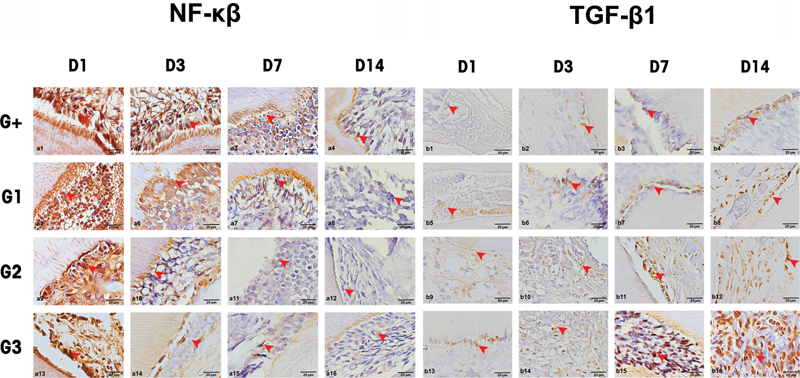
IHC results for NF-κB and TGF-β1 expression are presented in [Fig. 3]. One-way ANOVA results ([Table 1]) demonstrated significant differences in both markers across the four groups. For the proinflammatory marker NF-κB, significant variations were observed on days 1 and 3 (p < 0.05), indicating that the type of pulp capping material influenced the early inflammatory response. For the regenerative marker TGF-β1, significant differences were identified on days 1, 7, and 14 (p < 0.05), suggesting that material composition played a critical role in modulating reparative signaling over time.
Abbreviations: ANOVA, analysis of variance; NF-κB, nuclear factor-kappa β; SD, standard deviation; TGF-β1, transforming growth factor β 1.
Notes: Statistical analysis was performed using one-way ANOVA test. Exact p-values are presented; significance considered at p < 0.05. The values are mean ± SD.
Post hoc analysis ([Table 2]) further clarified specific intergroup differences. The positive control group (G + ), treated with 100% bioceramic material, exhibited significantly higher NF-κB expression on day 1 compared with all experimental groups containing calcium carbonate (G1, G2, and G3), indicating a more pronounced inflammatory reaction. Among the experimental groups, G3 demonstrated the lowest NF-κB expression on day 3, highlighting a stronger anti-inflammatory effect at this time point.
Abbreviations: NF-κB, nuclear factor-kappa β; SD, standard deviation; TGF-β1, transforming growth factor β 1ation.
Notes: Statistical analysis was performed using least-significance difference test. Exact p-values are presented; significance was considered at p < 0.05. The values are mean ± SD.
Regarding TGF-β1, post hoc comparisons showed that G2 and G3 consistently exhibited significantly higher expression levels compared with G+ and G1, particularly on days 7 and 14. These results suggest enhanced regenerative activity and anti-inflammatory signaling in groups treated with lower concentrations of calcium carbonate. The combination of calcium carbonate and bioceramic material in G2 and G3 appeared to modulate inflammation effectively while promoting tissue healing.
Temporal expression trends for both markers are illustrated in [Fig. 4], which shows a progressive decrease in NF-κB expression and a corresponding increase in TGF-β1 expression across all treatment groups throughout the 14-day observation period. This reciprocal pattern confirms the transition from an inflammatory to a regenerative state in this study.

Discussion
This study evaluated the biological response of rat dental pulp tissue to direct pulp capping using calcium carbonate synthesized from A. granosa shells combined with bioceramic material. The main findings demonstrated that experimental groups containing lower proportions of calcium carbonate exhibited significantly lower expression of the inflammatory marker NF-κB and significantly higher expression of the regenerative marker TGF-β1 compared with the bioceramic-only control. These results indicate that the addition of calcium carbonate effectively modulated inflammation while promoting reparative signaling in pulp tissue. Therefore, the findings of this study do not support the null hypothesis, as the incorporation of A. granosa shell-derived calcium carbonate significantly influenced both inflammatory and regenerative responses compared with bioceramic alone.
Anadara granosa shells are rich in calcium carbonate, a major inorganic constituent of bone and teeth. In dentistry, calcium carbonate has been utilized for bone grafting, remineralization, antibacterial action, and promoting wound healing through modulation of inflammation.[14]
This study observed peak NF-κB expression on days 1 and 3, consistent with findings by Hozhabri et al,[15] who reported that NF-κB is upregulated shortly after injury and diminishes as inflammation resolves. The early surge in NF-κB activates the release of inflammatory cytokines (e.g., TNF-α, IL-1β, IL-6, IL-8) and immune cells (neutrophils, macrophages, CD-47), potentially exacerbating neural inflammation and inducing apoptosis in pulpal cells if not regulated.[16]
A potential confounding factor in this study is the continuous eruption of rat incisors, which may influence early pulp responses through enhanced tissue turnover and mechanical stimulation of odontogenic cells. Such physiological activity could partly explain the elevated NF-κB levels observed at early time points (days 1–3), independent of material-induced effects. To minimize this confounder, all animals were age-matched (12–16 weeks) and treated using standardized cavity dimensions and exposure sites, while maintaining identical diet and housing conditions. Moreover, random allocation and blinded histological assessment were performed to reduce bias. The persistence of consistent expression trends at later time points (days 7 and 14), when eruptive activity tends to stabilize, suggests that the observed modulation of NF-κB and TGF-β1 was primarily material-driven rather than physiological.
TGF-β1 expression was elevated on days 7 and 14, aligning with its role in anti-inflammatory signaling and tissue regeneration. Lymphocytes and macrophages present during the chronic inflammatory phase secrete TGF-β1, which supports odontoblast-like cell differentiation and reparative dentin formation, indicating progression from inflammation toward healing.[17] These findings support the use of A. granosa-based calcium carbonate in modulating the early inflammatory response (via NF-κB) and enhancing regenerative signaling (via TGF-β1), contributing to favorable pulp healing outcomes.
The results showed that groups treated with calcium carbonate–bioceramic combinations exhibited a more substantial reduction in NF-κB expression compared with the bioceramic-only control group. This may be attributed to the release of calcium ions from calcium carbonate, which are known to regulate cellular signaling pathways and promote dental pulp cell differentiation. Calcium ions activate the ERK1/2 pathway, which inhibits IκB phosphorylation and suppresses NF-κB activity in proinflammatory cells, such as M1 macrophages, ultimately leading to reduced cytokine production and inflammation.[18]
These findings align with Saraswati et al,[19] who reported a reduction in NF-κB expression following pulp capping with calcium carbonate. Although calcium carbonate may cause mild inflammation, it does not induce necrosis in surrounding tissues, indicating good biocompatibility as reflected by the high reduction in NF-κB expression in the treatment groups. Among the experimental groups, G3 showed the greatest reduction in NF-κB expression. This effect may relate to the material's mechanical properties, as adequate compressive strength ensures structural stability of the capping agent under functional load.[20] Previous studies have indicated that high concentrations of calcium carbonate can reduce compressive strength due to CO2 release during mixing, potentially compromising material integrity.[21] [22] Additives such as propylene glycol or propolis have been proposed to counteract this effect and improve physical properties.[23]
This study also demonstrated increased expression of TGF-β1 in the calcium carbonate-treated groups, particularly on days 7 and 14. This can be attributed to the availability of calcium ions, which are critical for recruiting progenitor cells and promoting their proliferation and differentiation into odontoblast-like cells at the injury site.[24] These findings are consistent with those of Saraswati et al[17] and Kamba and Zakaria,[25] who observed that calcium carbonate derived from A. granosa enhances TGF-β1 expression. As inflammation subsides, anti-inflammatory and proregenerative signals become dominant, supported by the activation of M2 macrophages, which release growth factors including TGF-β1.[17]
The peak expression of TGF-β1 during the proliferative phase, typically between days 7 and 21, corresponds to fibroblast proliferation and collagen synthesis, both essential for tissue regeneration.[26] Although vascular endothelial growth factor-α was not the focus of this study, its known interaction with TGF-β1 in the regenerative cascade further supports the interpretation of enhanced healing in the calcium carbonate-treated groups.[27]
Statistical analysis confirmed that calcium carbonate improved the biological response to pulp capping treatment. Among all groups, G2 showed the most balanced outcome effectively controlling early inflammation while promoting TGF-β1-mediated tissue repair. G3 exhibited the highest TGF-β1 expression in later phases, indicating strong regenerative potential and suitability for long-term healing.
Throughout the study period, all animals remained clinically stable in terms of body weight around 10% threshold weight loss associated with moderate distress, and no animals reached the ≥20% terminal weight loss criterion that required humane euthanasia.[28] These observations are reinforced by IHC findings, which demonstrated favorable biological responses. Groups treated with lower proportions of calcium carbonate (G1 and G2) exhibited lower NF-κB expression and higher TGF-β1 levels, indicating effective modulation of inflammation and stimulation of tissue repair. These results support the local biocompatibility and regenerative potential of A. granosa-derived calcium carbonate as a functional additive in pulp capping materials.
One limitation of this study was the relatively short observation period, which may not fully capture the long-term dynamics of pulp healing and dentinal bridge formation. Future research should extend the evaluation period to approximately 28 or 56 days to monitor formation and maturation of the dentinal bridge. Incorporating advanced imaging modalities, such as micro-computed tomography, to assess dentinal bridge formation and further validate the safety and efficacy of A. granosa-based materials.
In addition, comparative analyses of cost and performance between A. granosa-derived calcium carbonate mixture and commercial bioceramic materials are warranted to establish their clinical and economic feasibility. Translational studies using larger animal models and eventually human clinical trials will be essential to validate the long-term safety, efficacy, and practical applicability of this sustainable biomaterial. Despite these limitations, the present study provides strong preliminary evidence supporting the therapeutic potential, cost efficiency, and environmental value of A. granosa-derived calcium carbonate as a bioactive and eco-friendly additive for enhancing the regenerative performance of bioceramic pulp capping agents.
Conclusion
Calcium carbonate synthesized from A. granosa shells, when combined with bioceramic material, demonstrated promising biocompatibility and regenerative potential as a direct pulp capping additive agent in vivo. Experimental formulations containing lower concentrations of calcium carbonate were associated with decreased NF-κB expression and elevated TGF-β1 expression, indicating effective modulation of the inflammatory response and enhanced reparative signaling in pulp tissue.
These results support the potential of natural calcium carbonate as a sustainable and bioactive additive to improve the therapeutic performance of bioceramic materials in vital pulp therapy. Future studies should include extended observation periods in larger animal models, followed by controlled clinical trials to confirm long-term safety, dentin bridge formation, and translational applicability in human patients.
Conflict of Interest
None declared.
Acknowledgments
We would like to thank the staffs of Biochemistry and Biomolecular Laboratory at Faculty of Medicine, Airlangga University for helping in IHC analysis.
-
References
- 1 Islam R, Islam MRR, Tanaka T, Alam MK, Ahmed HMA, Sano H. Direct pulp capping procedures - evidence and practice. Jpn Dent Sci Rev 2023; 59: 48-61
- 2 Saraswati W, Soetojo A, Dhaniar N. et al. CaCO3 from Anadara granosa shell as reparative dentin inducer in odontoblast pulp cells: in-vivo study. J Oral Biol Craniofac Res 2023; 13 (02) 164-168
- 3 Pribadi N, Widjiastuti I, Nadia A. Effect of calcium hydroxide-propolis combination on the number of fibroblast cells and angiogenesis in Wistar rats pulp. Conservative Dent J 2020; 10 (01) 14
- 4 Abdelwahab DH, Kabil NS, Badran AS, Darwish D, Abd El Geleel OM. One-year radiographic and clinical performance of bioactive materials in primary molar pulpotomy: a randomized controlled trial. J Dent 2024; 143: 104864
- 5 Davaie S, Hooshmand T, Ansarifard S. Different types of bioceramics as dental pulp capping materials: a systematic review. Ceram Int 2021; 47 (15) 20781-20792
- 6 Chae YK, Ye JR, Nam OH. Evaluation of biocompatibility and bioactive potential of Well-Root PT by comparison with ProRoot MTA and Biodentine. J Dent Sci 2024; 19 (04) 2218-2225
- 7 Dhanaraj K, Suresh G. Conversion of waste sea shell (Anadara granosa) into valuable nanohydroxyapatite (nHAp) for biomedical applications. Vacuum 2018; 152: 222-230
- 8 Cheng M, Liu M, Chang L. et al. Overview of structure, function and integrated utilization of marine shell. Sci Total Environ 2023; 870: 161950
- 9 Muntean FL, Olariu I, Marian D. et al. Hydroxyapatite from Mollusk shells: characteristics, production, and potential applications in dentistry. Dent J 2024; 12 (12) 409
- 10 Cunha Nde O, Junqueira MA, Cosme-Silva L. et al. Expression of matrix metalloproteinases-8 and myeloperoxidase in pulp tissue after pulpotomy with calcium silicate cements. Pesqui Bras Odontopediatria Clin Integr. 2021; 21
- 11 Boopathi T, Manimaran S, Kerena JC, Sebeena M, Karthick K, Deepa NT. Histological evaluation of pulp response to alendronate and Biodentine as pulp capping agents: an animal study. Restor Dent Endod 2024; 49 (04) e39
- 12 Elmubarak NA. The road map to proper dental pulp experiments in animal models. Int Dent J Stud Res 2024; 11 (04) 163-169
- 13 Percie du Sert N, Hurst V, Ahluwalia A. et al. The ARRIVE guidelines 2.0: updated guidelines for reporting animal research. PLoS Biol 2020; 18 (07) e3000410
- 14 Asmawati A, Thalib B, Thalib AM, Reni DS, Hasyim R. Comparison of blood clam (Anadara granosa) shell paste, shrimp (Litopenaeus vannamei) shell paste and casein phosphopeptide-amorphus calcium phosphate (CPP-ACP) paste as teeth remineralization material. J Dentomaxillofac Sci 2018; 3 (03) 162
- 15 Hozhabri NST, Benson MD, Vu MD. et al. Decreasing NF-κB expression enhances odontoblastic differentiation and collagen expression in dental pulp stem cells exposed to inflammatory cytokines. PLoS One 2015; 10 (01) e0113334
- 16 Vaseenon S, Weekate K, Srisuwan T, Chattipakorn N, Chattipakorn S. Observation of inflammation, oxidative stress, mitochondrial dynamics, and apoptosis in dental pulp following a diagnosis of irreversible pulpitis. Eur Endod J 2023; 8 (02) 148-155
- 17 Saraswati W, Yahya AN, Yonas Y. et al. Calcium carbonate from Anadara granosa shells stimulates FGF2, TGF-β1, and collagen type 1 expression in rat dental pulp. Eur J Dent 2025; 19 (03) 811-816
- 18 Landén NX, Li D, Ståhle M. Transition from inflammation to proliferation: a critical step during wound healing. Cell Mol Life Sci 2016; 73 (20) 3861-3885
- 19 Saraswati W, Dhaniar N, Wahjuningrum DA, Nuraini N, Bhardwaj A. The Effect of Exposure calcium carbonate from blood cockle (Anadara granosa) shells to the expression of the NF-κβ on dentin pulp complex. J Int Dent Med Res 2021; 14 (02) 549-553
- 20 Galić VO, Stamenić Z, Petrović V, Jokanović V, Živković S. Compressive strength of calcium silicate-based cement. Stomatol Glas Srb 2018; 65 (01) 7-13
- 21 Saraswati W, Juniarti DE, Lestari V. et al. Differences of compressive strength between calcium carbonate from blood clam shells and calcium hydroxide as a candidate for pulp capping material. Conservative Dent J 2024; 14 (01) 11-14
- 22 Bernardi A, Bortoluzzi EA, Felippe WT, Felippe MCS, Wan WS, Teixeira CS. Effects of the addition of nanoparticulate calcium carbonate on setting time, dimensional change, compressive strength, solubility and pH of MTA. Int Endod J 2017; 50 (01) 97-105
- 23 Widjiastuti I, Setyabudi S, Mudjiono M, Setyowati E. Compressive strength test on calcium hydroxide with propolis combination. Conservative Dent J 2019; 9 (01) 28
- 24 Juniarti DE, Kunarti S, Mardiyah AA, Purniati NMD. Biomodulator of diode laser irradiation on odontoblast-like cells by expression of vascular endothelial growth factor-A and transforming growth factor-β1. Eur J Dent 2023; 17 (03) 706-712
- 25 Shafiu Kamba A, Zakaria ZAB. Osteoblasts growth behaviour on bio-based calcium carbonate aragonite nanocrystal. BioMed Res Int 2014; 2014: 215097
- 26 Mustakim KR, Eo MY, Seo MH. et al. Ultrastructural and immunohistochemical evaluation of hyperplastic soft tissues surrounding dental implants in fibular jaws. Sci Rep 2024; 14 (01) 10717
- 27 Sularsih S, Fransiska W, Salsabila S, Rahmitasari F, Soesilo D, Prananingrum W. Potency of the combination of chitosan and hydroxyapatite on angiogenesis and fibroblast cell proliferation in direct pulp capping of Rattus norvegicus . Eur J Dent 2024; 18 (04) 1135-1141
- 28 Texas A&M University Division of Research. Guidelines on Choosing Appropriate Endpoints (TAMU-G-001). 2024 . Accessed July 23, 2025 at: https://research.tamu.edu/wp-content/uploads/2024/12/TAMU-G-001-IACUC-Guidelines-on-Choosing-Appropriate-Endpoints-1.pdf
Address for correspondence
Publication History
Article published online:
22 January 2026
© 2026. The Author(s). This is an open access article published by Thieme under the terms of the Creative Commons Attribution License, permitting unrestricted use, distribution, and reproduction so long as the original work is properly cited. (https://creativecommons.org/licenses/by/4.0/)
Thieme Medical and Scientific Publishers Pvt. Ltd.
A-12, 2nd Floor, Sector 2, Noida-201301 UP, India
-
References
- 1 Islam R, Islam MRR, Tanaka T, Alam MK, Ahmed HMA, Sano H. Direct pulp capping procedures - evidence and practice. Jpn Dent Sci Rev 2023; 59: 48-61
- 2 Saraswati W, Soetojo A, Dhaniar N. et al. CaCO3 from Anadara granosa shell as reparative dentin inducer in odontoblast pulp cells: in-vivo study. J Oral Biol Craniofac Res 2023; 13 (02) 164-168
- 3 Pribadi N, Widjiastuti I, Nadia A. Effect of calcium hydroxide-propolis combination on the number of fibroblast cells and angiogenesis in Wistar rats pulp. Conservative Dent J 2020; 10 (01) 14
- 4 Abdelwahab DH, Kabil NS, Badran AS, Darwish D, Abd El Geleel OM. One-year radiographic and clinical performance of bioactive materials in primary molar pulpotomy: a randomized controlled trial. J Dent 2024; 143: 104864
- 5 Davaie S, Hooshmand T, Ansarifard S. Different types of bioceramics as dental pulp capping materials: a systematic review. Ceram Int 2021; 47 (15) 20781-20792
- 6 Chae YK, Ye JR, Nam OH. Evaluation of biocompatibility and bioactive potential of Well-Root PT by comparison with ProRoot MTA and Biodentine. J Dent Sci 2024; 19 (04) 2218-2225
- 7 Dhanaraj K, Suresh G. Conversion of waste sea shell (Anadara granosa) into valuable nanohydroxyapatite (nHAp) for biomedical applications. Vacuum 2018; 152: 222-230
- 8 Cheng M, Liu M, Chang L. et al. Overview of structure, function and integrated utilization of marine shell. Sci Total Environ 2023; 870: 161950
- 9 Muntean FL, Olariu I, Marian D. et al. Hydroxyapatite from Mollusk shells: characteristics, production, and potential applications in dentistry. Dent J 2024; 12 (12) 409
- 10 Cunha Nde O, Junqueira MA, Cosme-Silva L. et al. Expression of matrix metalloproteinases-8 and myeloperoxidase in pulp tissue after pulpotomy with calcium silicate cements. Pesqui Bras Odontopediatria Clin Integr. 2021; 21
- 11 Boopathi T, Manimaran S, Kerena JC, Sebeena M, Karthick K, Deepa NT. Histological evaluation of pulp response to alendronate and Biodentine as pulp capping agents: an animal study. Restor Dent Endod 2024; 49 (04) e39
- 12 Elmubarak NA. The road map to proper dental pulp experiments in animal models. Int Dent J Stud Res 2024; 11 (04) 163-169
- 13 Percie du Sert N, Hurst V, Ahluwalia A. et al. The ARRIVE guidelines 2.0: updated guidelines for reporting animal research. PLoS Biol 2020; 18 (07) e3000410
- 14 Asmawati A, Thalib B, Thalib AM, Reni DS, Hasyim R. Comparison of blood clam (Anadara granosa) shell paste, shrimp (Litopenaeus vannamei) shell paste and casein phosphopeptide-amorphus calcium phosphate (CPP-ACP) paste as teeth remineralization material. J Dentomaxillofac Sci 2018; 3 (03) 162
- 15 Hozhabri NST, Benson MD, Vu MD. et al. Decreasing NF-κB expression enhances odontoblastic differentiation and collagen expression in dental pulp stem cells exposed to inflammatory cytokines. PLoS One 2015; 10 (01) e0113334
- 16 Vaseenon S, Weekate K, Srisuwan T, Chattipakorn N, Chattipakorn S. Observation of inflammation, oxidative stress, mitochondrial dynamics, and apoptosis in dental pulp following a diagnosis of irreversible pulpitis. Eur Endod J 2023; 8 (02) 148-155
- 17 Saraswati W, Yahya AN, Yonas Y. et al. Calcium carbonate from Anadara granosa shells stimulates FGF2, TGF-β1, and collagen type 1 expression in rat dental pulp. Eur J Dent 2025; 19 (03) 811-816
- 18 Landén NX, Li D, Ståhle M. Transition from inflammation to proliferation: a critical step during wound healing. Cell Mol Life Sci 2016; 73 (20) 3861-3885
- 19 Saraswati W, Dhaniar N, Wahjuningrum DA, Nuraini N, Bhardwaj A. The Effect of Exposure calcium carbonate from blood cockle (Anadara granosa) shells to the expression of the NF-κβ on dentin pulp complex. J Int Dent Med Res 2021; 14 (02) 549-553
- 20 Galić VO, Stamenić Z, Petrović V, Jokanović V, Živković S. Compressive strength of calcium silicate-based cement. Stomatol Glas Srb 2018; 65 (01) 7-13
- 21 Saraswati W, Juniarti DE, Lestari V. et al. Differences of compressive strength between calcium carbonate from blood clam shells and calcium hydroxide as a candidate for pulp capping material. Conservative Dent J 2024; 14 (01) 11-14
- 22 Bernardi A, Bortoluzzi EA, Felippe WT, Felippe MCS, Wan WS, Teixeira CS. Effects of the addition of nanoparticulate calcium carbonate on setting time, dimensional change, compressive strength, solubility and pH of MTA. Int Endod J 2017; 50 (01) 97-105
- 23 Widjiastuti I, Setyabudi S, Mudjiono M, Setyowati E. Compressive strength test on calcium hydroxide with propolis combination. Conservative Dent J 2019; 9 (01) 28
- 24 Juniarti DE, Kunarti S, Mardiyah AA, Purniati NMD. Biomodulator of diode laser irradiation on odontoblast-like cells by expression of vascular endothelial growth factor-A and transforming growth factor-β1. Eur J Dent 2023; 17 (03) 706-712
- 25 Shafiu Kamba A, Zakaria ZAB. Osteoblasts growth behaviour on bio-based calcium carbonate aragonite nanocrystal. BioMed Res Int 2014; 2014: 215097
- 26 Mustakim KR, Eo MY, Seo MH. et al. Ultrastructural and immunohistochemical evaluation of hyperplastic soft tissues surrounding dental implants in fibular jaws. Sci Rep 2024; 14 (01) 10717
- 27 Sularsih S, Fransiska W, Salsabila S, Rahmitasari F, Soesilo D, Prananingrum W. Potency of the combination of chitosan and hydroxyapatite on angiogenesis and fibroblast cell proliferation in direct pulp capping of Rattus norvegicus . Eur J Dent 2024; 18 (04) 1135-1141
- 28 Texas A&M University Division of Research. Guidelines on Choosing Appropriate Endpoints (TAMU-G-001). 2024 . Accessed July 23, 2025 at: https://research.tamu.edu/wp-content/uploads/2024/12/TAMU-G-001-IACUC-Guidelines-on-Choosing-Appropriate-Endpoints-1.pdf